File:Meyer1914 fig03.jpg
From Embryology

Size of this preview: 623 × 600 pixels. Other resolution: 831 × 800 pixels.
Original file (831 × 800 pixels, file size: 69 KB, MIME type: image/jpeg)
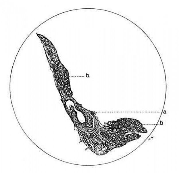
Fig. 3 Drawing of the unrolled border of an accessory free fold from the suspensory ligament to the gall bladder of the dog
The large thick-walled vein is shown at a point where one of its branches joins. a, vein; 1), fat. X42.
| Historic Disclaimer - information about historic embryology pages |
|---|
 |
Reference
Cite this page: Hill, M.A. (2024, April 27) Embryology Meyer1914 fig03.jpg. Retrieved from https://embryology.med.unsw.edu.au/embryology/index.php/File:Meyer1914_fig03.jpg
- © Dr Mark Hill 2024, UNSW Embryology ISBN: 978 0 7334 2609 4 - UNSW CRICOS Provider Code No. 00098G
File history
Click on a date/time to view the file as it appeared at that time.
| Date/Time | Thumbnail | Dimensions | User | Comment | |
|---|---|---|---|---|---|
| current | 21:06, 3 November 2015 | | 831 × 800 (69 KB) | Z8600021 (talk | contribs) | |
| 21:06, 3 November 2015 |  | 1,324 × 1,008 (127 KB) | Z8600021 (talk | contribs) | Fig. 3 Drawing of the unrolled border of an accessory free fold from the suspensory ligament to the gall bladder of the dog. The large thick-walled vein is shown at a point where one of its branches joins. a, vein; 1), fat. X42. |
You cannot overwrite this file.
File usage
The following page uses this file: