File:Menstrual cycle - brain subcortical structural changes.png

Original file (900 × 390 pixels, file size: 134 KB, MIME type: image/png)
Subcortical structural changes along the menstrual cycle
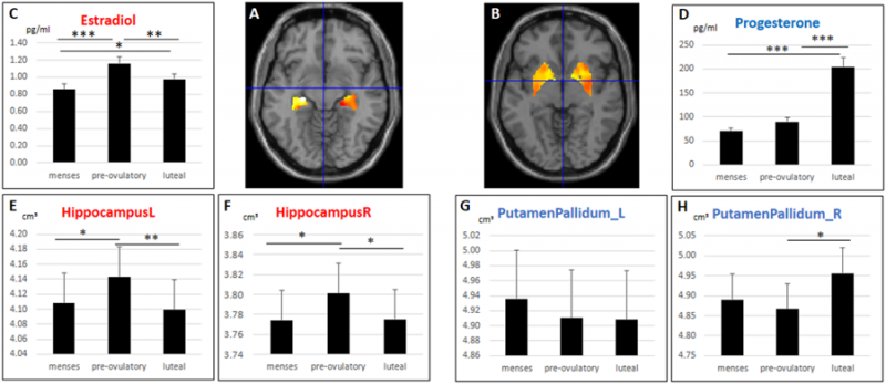
Gray matter volumes extracted from the left and right hippocampus were significantly larger in the pre-ovulatory phase, when estradiol levels peak, compared to menses and luteal cycle phase. The effect was indeed driven by estradiol levels. Gray matter volumes extracted from the right basal ganglia were significantly larger in the luteal phase, when progesterone levels peak, compared to the pre-ovulatory phase. The effect was indeed driven by progesterone levels. (A) Hippocampal regions of interest, (B) Basal ganglia regions of interest, (C) Estradiol levels during the three cycle phases, (D) Progesterone levels during the three cycle phases, (E,F) Left and right hippocampal volumes during the three cycle phases, (G,H): left and right basal ganglia volumes during the three cycle phases. *p < 0.05, **p < 0.01, ***p < 0.001. Error bars represent standard errors.
Reference
Pletzer B, Harris T & Hidalgo-Lopez E. (2018). Subcortical structural changes along the menstrual cycle: beyond the hippocampus. Sci Rep , 8, 16042. PMID: 30375425 DOI.
Copyright
Open Access This article is licensed under a Creative Commons Attribution 4.0 International License, which permits use, sharing, adaptation, distribution and reproduction in any medium or format, as long as you give appropriate credit to the original author(s) and the source, provide a link to the Creative Commons license, and indicate if changes were made. The images or other third party material in this article are included in the article’s Creative Commons license, unless indicated otherwise in a credit line to the material. If material is not included in the article’s Creative Commons license and your intended use is not permitted by statutory regulation or exceeds the permitted use, you will need to obtain permission directly from the copyright holder. To view a copy of this license, visit http://creativecommons.org/licenses/by/4.0/.
41598_2018_34247_Fig1_HTML.png
File history
Click on a date/time to view the file as it appeared at that time.
| Date/Time | Thumbnail | Dimensions | User | Comment | |
|---|---|---|---|---|---|
| current | 12:03, 6 November 2018 |  | 900 × 390 (134 KB) | Z8600021 (talk | contribs) | ==Subcortical structural changes along the menstrual cycle== Gray matter volumes extracted from the left and right hippocampus were significantly larger in the pre-ovulatory phase, when estradiol levels peak, compared to menses and luteal cycle phase.... |
You cannot overwrite this file.
File usage
The following page uses this file: