File:Melanocytes in Eyes of B6 and ep Mice.jpeg

Original file (513 × 634 pixels, file size: 112 KB, MIME type: image/jpeg)
Figure 2.
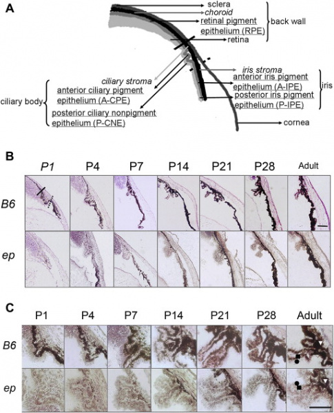
Pigmentation of the anterior segment of the eyes in B6 and ep mice. (A) Sagittal diagram of anterior eye of adult mouse. Tissues containing melanocytes derived from the neural crest were italicised and pigmented cells derived from the neuroepithelium of the optic cup were underlined. (B) Pigmentation of the anterior segment of the eyes of B6 and ep mice. As shown in the upper-left photo, the iris is defined as the part of tunica vascularis beneath the dotted line; ciliary body is defined as the part between solid and dotted lines; choroid/retina is defined as the part above the solid line. The bar indicates 100 mm. (C) Pigmentation of the ciliary body and ciliary storm in postnatal days, adult, and 3 months old. The bar indicates 50 mm.
! 2013 Elsevier Ltd. All rights reserved.
File history
Click on a date/time to view the file as it appeared at that time.
| Date/Time | Thumbnail | Dimensions | User | Comment | |
|---|---|---|---|---|---|
| current | 17:28, 4 September 2018 |  | 513 × 634 (112 KB) | Z5165679 (talk | contribs) | Pigmentation of the anterior segment of the eyes in B6 and ep mice. (A) Sagittal diagram of anterior eye of adult mouse. Tissues containing melanocytes derived from the neural crest were italicised and pigmented cells derived from the neuroepithelium o... |
You cannot overwrite this file.
File usage
The following 3 pages use this file: