File:McMurrich1930 fig85.jpg: Difference between revisions
(Z8600021 uploaded a new version of File:McMurrich1930 fig85.jpg) |
mNo edit summary |
||
| (One intermediate revision by the same user not shown) | |||
| Line 1: | Line 1: | ||
== | ==Fig. 85. Representations of human fetus at term and of ungulate placenta== | ||
Fig. 85. Representations of human fetus at term and of ungulate placenta | |||
(QIII, 8.) | |||
So too it is the chorion of the fetal calf that is described, and it was upon this that Leonardo, following the example of his predecessors, based his ideas of human placentation, assuming that it also was of the cotyledonary type. In the wonderful drawing (fig. 85) of the fetus still within the womb (QIII, 8) it is curious to note cotyledons of the ungulate type on the wall of the uterus, while the discoidal placenta is entirely overlooked, even although there is a memorandum to “Note well the umbilical vein where it ends in the uterus” (QIII, 7v). It is evident that Leonardo had examined a pregnant human uterus at term— he gives a number of drawings of the fetus as it lies curled up within the womb (QIII, 7, 7v, 8, 9v) — but in none of them is a placenta shown. Where the uterus is also shown there is usually no indication of how the fetus is connected with it; only in QIII, 8 are the cotyledons represented, on the assumption that what he had seen in the cow occurred also in the human pregnant uterus. | |||
===Reference=== | ===Reference=== | ||
Latest revision as of 11:46, 20 April 2020
Fig. 85. Representations of human fetus at term and of ungulate placenta
(QIII, 8.)
So too it is the chorion of the fetal calf that is described, and it was upon this that Leonardo, following the example of his predecessors, based his ideas of human placentation, assuming that it also was of the cotyledonary type. In the wonderful drawing (fig. 85) of the fetus still within the womb (QIII, 8) it is curious to note cotyledons of the ungulate type on the wall of the uterus, while the discoidal placenta is entirely overlooked, even although there is a memorandum to “Note well the umbilical vein where it ends in the uterus” (QIII, 7v). It is evident that Leonardo had examined a pregnant human uterus at term— he gives a number of drawings of the fetus as it lies curled up within the womb (QIII, 7, 7v, 8, 9v) — but in none of them is a placenta shown. Where the uterus is also shown there is usually no indication of how the fetus is connected with it; only in QIII, 8 are the cotyledons represented, on the assumption that what he had seen in the cow occurred also in the human pregnant uterus.
Reference
McMurrich JP. Leonardo da Vinci - the anatomist. (1930) Carnegie institution of Washington, Williams & Wilkins Company, Baltimore.
Cite this page: Hill, M.A. (2024, May 21) Embryology McMurrich1930 fig85.jpg. Retrieved from https://embryology.med.unsw.edu.au/embryology/index.php/File:McMurrich1930_fig85.jpg
- © Dr Mark Hill 2024, UNSW Embryology ISBN: 978 0 7334 2609 4 - UNSW CRICOS Provider Code No. 00098G
File history
Click on a date/time to view the file as it appeared at that time.
| Date/Time | Thumbnail | Dimensions | User | Comment | |
|---|---|---|---|---|---|
| current | 09:48, 25 March 2020 | 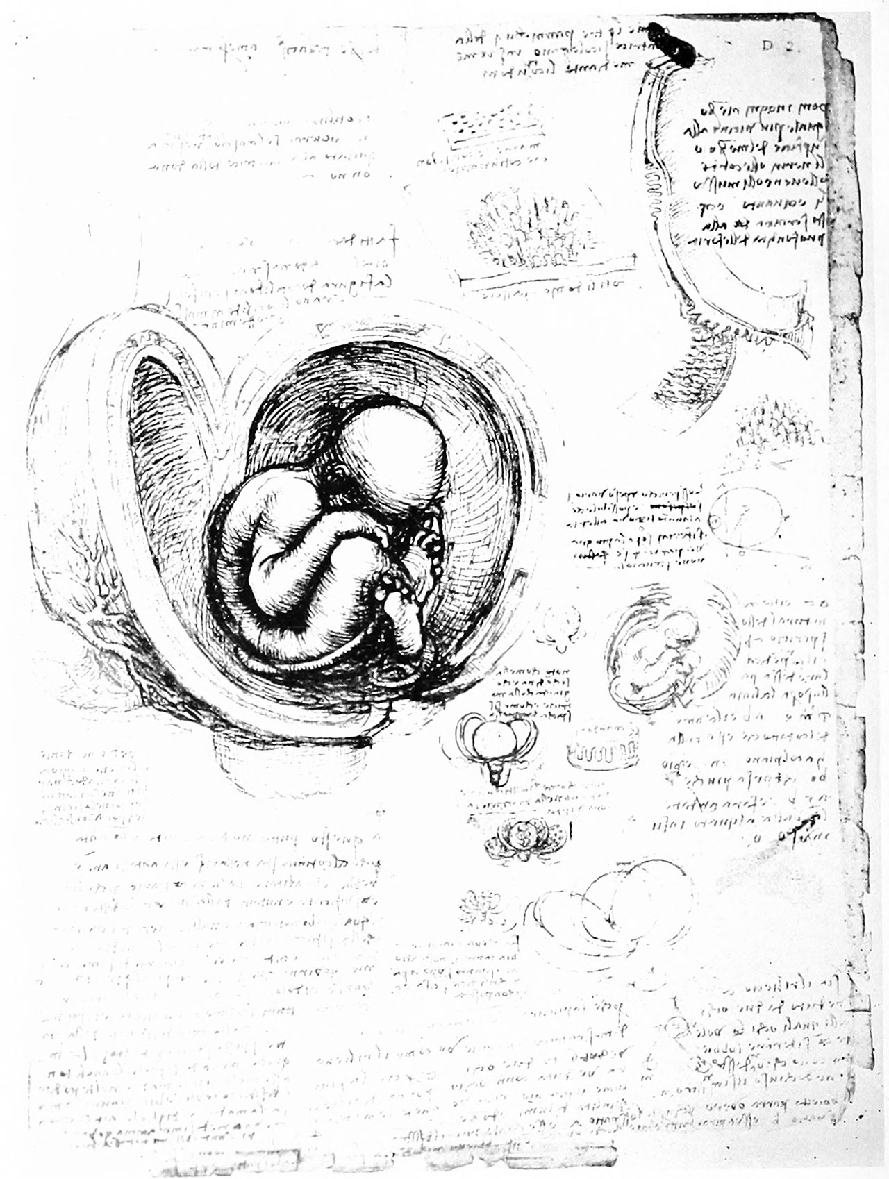 | 1,280 × 1,698 (373 KB) | Z8600021 (talk | contribs) | BW and scaled to 1280 pixels wide |
| 09:48, 25 March 2020 | 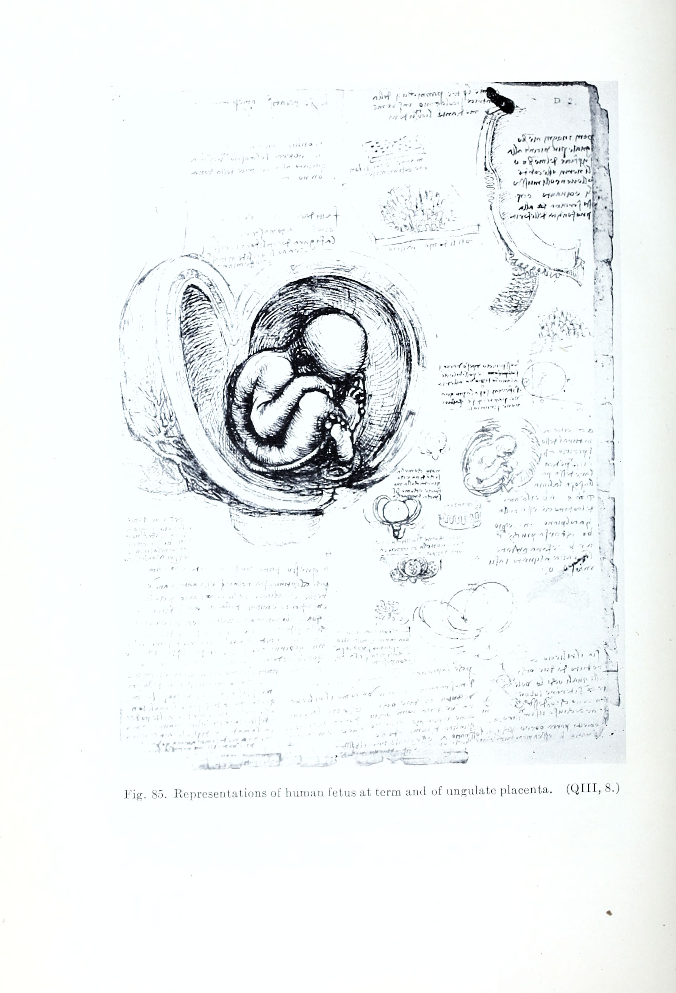 | 2,224 × 3,266 (594 KB) | Z8600021 (talk | contribs) | Fig. 85. Representations of human fetus at term and of ungulate placenta. (QIII, 8.) ===Reference=== {{Ref-McMurrich1930}} {{Footer}} Category:Historic EmbryologyCategory:FetalCategory:Placenta |
You cannot overwrite this file.
File usage
The following 2 pages use this file: