File:McMurrich1930 fig84.jpg

Original file (1,280 × 1,819 pixels, file size: 784 KB, MIME type: image/jpeg)
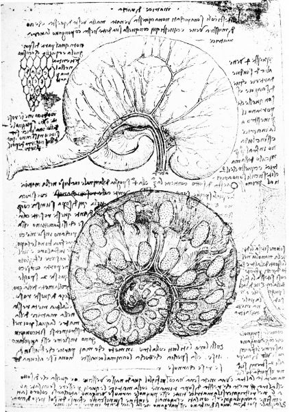
Fig. 84. Two figures of membranes and circulation of fetal calf
(AnB, 28.)
The representation of these last two investments indicates that the sketch is intended as a diagram of the membranes surrounding a mammalian embryo, and two excellent figures on AnB, 38 (fig. 84) show that Leonardo had made a study of the pregnant uterus of a cow. In one figure he shows such a uterus with one of the ovaries and four blood-vessels supplying its walls, “these four veins a, b, c, d are tw T o of arteries and two of blood,” while the cotyledons show through indistinctly. In the second figure the fetus is shown still enclosed within the chorion, over whose surface the cotyledons, now plainly seen, are scattered and blood-vessels ramify in it, coming from the umbilical cord which is seen issuing from the ventral abdominal wall of the fetus.
Reference
McMurrich JP. Leonardo da Vinci - the anatomist. (1930) Carnegie institution of Washington, Williams & Wilkins Company, Baltimore.
Cite this page: Hill, M.A. (2024, April 27) Embryology McMurrich1930 fig84.jpg. Retrieved from https://embryology.med.unsw.edu.au/embryology/index.php/File:McMurrich1930_fig84.jpg
- © Dr Mark Hill 2024, UNSW Embryology ISBN: 978 0 7334 2609 4 - UNSW CRICOS Provider Code No. 00098G
File history
Click on a date/time to view the file as it appeared at that time.
| Date/Time | Thumbnail | Dimensions | User | Comment | |
|---|---|---|---|---|---|
| current | 09:44, 25 March 2020 |  | 1,280 × 1,819 (784 KB) | Z8600021 (talk | contribs) | BW and scaled to 1280 pixels wide |
| 09:43, 25 March 2020 |  | 2,264 × 3,279 (1.11 MB) | Z8600021 (talk | contribs) | Fig. 84. Two figures of membranes and circulation of fetal calf. (AnB, 28.) ===Reference=== {{Ref-McMurrich1930}} {{Footer}} |
You cannot overwrite this file.
File usage
The following 2 pages use this file: