File:MartinFalkiner1938 plate02.jpg

Original file (1,280 × 1,840 pixels, file size: 247 KB, MIME type: image/jpeg)
Plate 2
5 Section 30 B. X150. This section passes through the primitive streak, which is seen at the caudal half of the embryonic plate. The amnion and embryonie plate have been torn. A t0ng'ue of tissue still pa.1'tia.lly divides the yolk sac.
6 Section 30 E. X 150. The amnion and embryonic plate are much damaged but the latter is now sectioned at approximately 90° and is seen to be composed of a densely packed columnar epithelium. The roof of the yolk sac is formed by an even rod of tissue, two or more cells in thickness but thinning out at the cephalic end. This is interpreted as the head process or notoehordal plate.
7 Section 3.1. D. X 150. The embryonic plate is now cut through at 90°. The amnion has rapidly diminislled in size. A loose Strand of mesoderm intervenes between the embryonic plate and the underlying endoderrn. The significance of the detached clump of cells in the middle of the yolk see is unknown.
8 Reconstruction of medial plane of embryo.
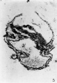
Fig. 5
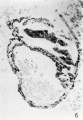
Fig. 6
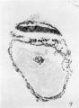
Fig. 7
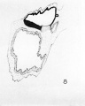
Fig. 8


Reference
Martin CP. and Falkiner N. Mcl. The Falkiner ovum. (1938) Amer. J Anat., 63: 251-271.
Cite this page: Hill, M.A. (2024, April 27) Embryology MartinFalkiner1938 plate02.jpg. Retrieved from https://embryology.med.unsw.edu.au/embryology/index.php/File:MartinFalkiner1938_plate02.jpg
- © Dr Mark Hill 2024, UNSW Embryology ISBN: 978 0 7334 2609 4 - UNSW CRICOS Provider Code No. 00098G
File history
Click on a date/time to view the file as it appeared at that time.
| Date/Time | Thumbnail | Dimensions | User | Comment | |
|---|---|---|---|---|---|
| current | 11:57, 11 August 2017 | | 1,280 × 1,840 (247 KB) | Z8600021 (talk | contribs) | |
| 11:56, 11 August 2017 |  | 1,649 × 2,388 (325 KB) | Z8600021 (talk | contribs) |
You cannot overwrite this file.
File usage
The following page uses this file: