File:MartinFalkiner1938 fig03.jpg

Original file (700 × 1,000 pixels, file size: 89 KB, MIME type: image/jpeg)
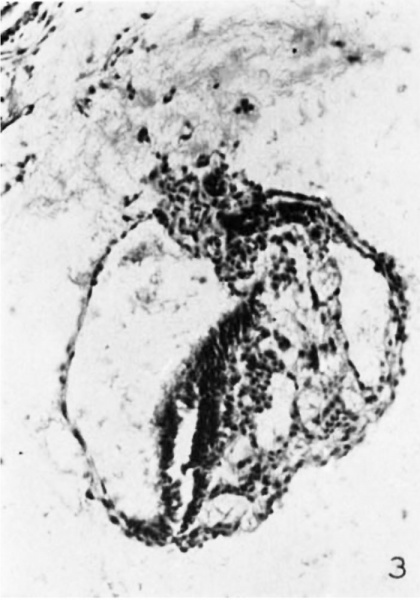
Fig. 3
Section 29 D. X 150. Two parts of the yolk sac cavity separated by a broad band of tissue are visible in this section. One small part lies fairly close under the embryonic plate; the other and larger lies more ventrally. This last is the compartment that was visible in previous sections. The allantois is cut in two places: first it is seen as a long duct running dorsally from the caudal end of the ventral compartment of the yolk sac, and secondly it is again c.ut slightly caudal to the dorsal end of the duct-like part. From the dorsal end of the longitudinal part a wedge-shaped mass of cells projects toward the amniotic cavity.
Reference
Martin CP. and Falkiner N. Mcl. The Falkiner ovum. (1938) Amer. J Anat., 63: 251-271.
Cite this page: Hill, M.A. (2024, April 27) Embryology MartinFalkiner1938 fig03.jpg. Retrieved from https://embryology.med.unsw.edu.au/embryology/index.php/File:MartinFalkiner1938_fig03.jpg
- © Dr Mark Hill 2024, UNSW Embryology ISBN: 978 0 7334 2609 4 - UNSW CRICOS Provider Code No. 00098G
File history
Click on a date/time to view the file as it appeared at that time.
| Date/Time | Thumbnail | Dimensions | User | Comment | |
|---|---|---|---|---|---|
| current | 11:46, 11 August 2017 |  | 700 × 1,000 (89 KB) | Z8600021 (talk | contribs) |
You cannot overwrite this file.
File usage
The following 3 pages use this file: