File:Marshall1881 plate01.jpg
Original file (941 × 1,493 pixels, file size: 455 KB, MIME type: image/jpeg)
Explanation of Plate I
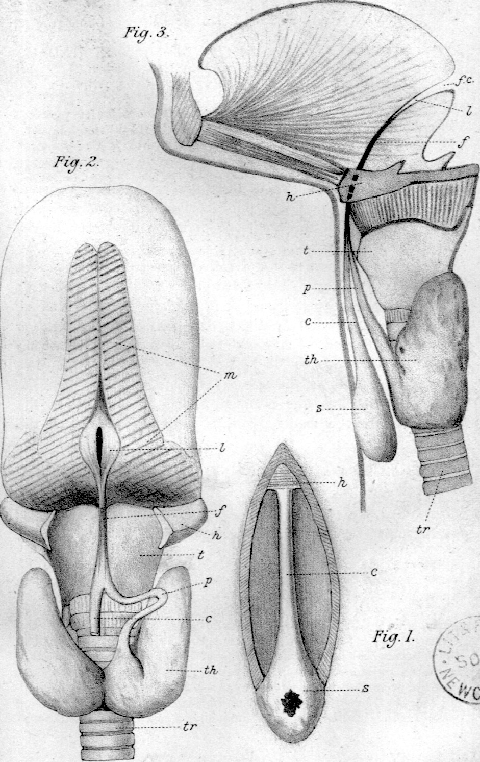
Fig. 1 shows the canal and sac exposed by reflecting the skin from the middle line of the neck.
Fig. 2 shows a dissection of the thyroid gland and under surface of the tongue. The lobus pyramidalis is seen arising from the left side of the thyroid gland; the upper end of the canal is joined by the pyramid, and the two structures are continued onwards as the median fibrous cord, which is exposed by dividing the hyoid bone. Further on the canal is seen to become patent and pass up to the foramen cecum.
Fig. 3 is a slightly diagrammatic view of the parts in question. The tongue and upper canal (lingual duct) are shown in vertical section as far down as the hyoid bone; below the hyoid bone the parts are seen in elevation. The thyro-glossal duct is here shown in its whole length, the shaded portion representing the obliterated part which I have referred to as the fibrous cord.
Plate I
(All the figures are natural size.)
Fig. 1. Canal of His exposed by dissecting the skin from the median line of the neck. c¢, canal; 2, hyoid bone; s, sac with open sinus.
Fig. 2. Dissection of the thyro-glossal duct, thyroid gland, and base of the tongue. , upper end of canal of His; f, fibrous cord; A, hyoid bone; J, lingual duct opened and fixed by pins; m, muscles cut through at base of tongue; p, pyramidal lobe; z, thyroid cartilage; ¢h, thyroid gland ; tr, trachea. ;
Fig. 3. Semi-diagrammatic figure: the upper parts are in vertical section, the lower part in elevation; /f. c, foramen cecum; s, sac with sinus opening in front of neck ; other letters as before.
Reference
Marshall AM. Thyro-Glossal duct or “canal of His”. (1881) J Anat. Physiol. 16(3): 305–354. PMID 17231961
Cite this page: Hill, M.A. (2024, April 27) Embryology Marshall1881 plate01.jpg. Retrieved from https://embryology.med.unsw.edu.au/embryology/index.php/File:Marshall1881_plate01.jpg
- © Dr Mark Hill 2024, UNSW Embryology ISBN: 978 0 7334 2609 4 - UNSW CRICOS Provider Code No. 00098G
File history
Click on a date/time to view the file as it appeared at that time.
| Date/Time | Thumbnail | Dimensions | User | Comment | |
|---|---|---|---|---|---|
| current | 09:27, 22 July 2020 | | 941 × 1,493 (455 KB) | Z8600021 (talk | contribs) | |
| 09:24, 22 July 2020 |  | 1,177 × 1,626 (415 KB) | Z8600021 (talk | contribs) | ==Explanation of Plate I== (All the figures are natural size.) Fig. 1. Canal of His exposed by dissecting the skin from the median line of the neck. c¢, canal; 2, hyoid bone; s, sac with open sinus. Fig. 2. Dissection of the thyro-glossal duct, thyroid gland, and base of the tongue. , upper end of canal of His; f, fibrous cord; A, hyoid bone; J, lingual duct opened and fixed by pins; m, muscles cut through at base of tongue; p, pyramidal lobe; z, thyroid cartilage; ¢h, thyroid gland ; tr,... |
You cannot overwrite this file.
File usage
The following page uses this file: