File:Marshall1879 fig22.jpg
From Embryology

Size of this preview: 800 × 509 pixels. Other resolution: 819 × 521 pixels.
Original file (819 × 521 pixels, file size: 110 KB, MIME type: image/jpeg)
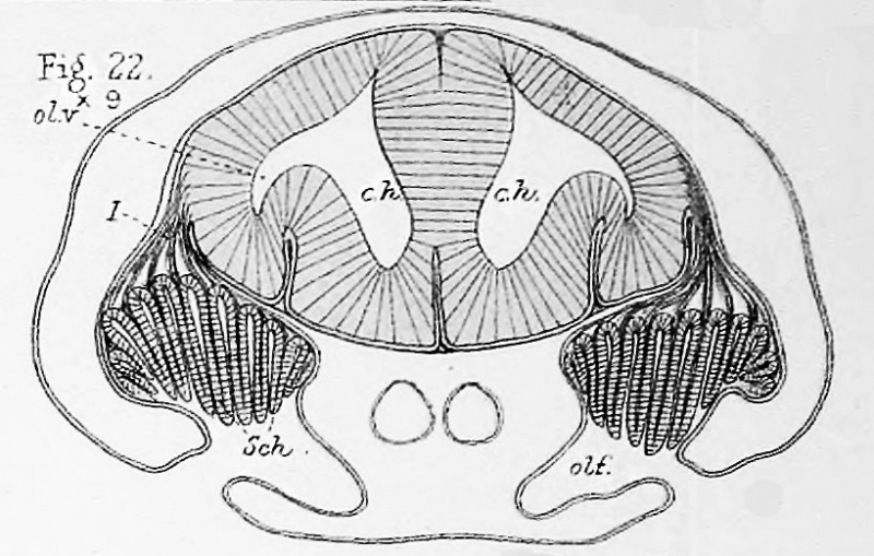
Fig. 22. Transverse section through the forepart of the head of a dogfish embryo of stage Q
Showing the cerebral hemispheres, olfactory lobes, olfactory nerves, and olfactory pits with their Schneiderian folds, x 9 diam.
| Alphabetical List of References |
|---|
| ah. Alimentary canal. aV. Anterior prolongation of alimentary canal. ao.r Dorsal aorta. a. r. Anterior root of a spinal nerve. mtcl. Auditory vesicle. b. a. Branchial artery. br. 1. First branchial arch. br. 2. Second branchial arch. c. h. cerebral hemisphere. f. b. forebrain, g. gill. h. 2. Second head-cavity. h . b. Hindbrain. hy. Hyoid arch. inf. Infundibulum, I. c. Lachrymal cleft. m. b. Midbrain. Mn. Mandibular arch. m. p. Muscle plate. M.v. Maxillary arch. n. Notochord. o. c. Optic cup or eye. olf. Olfactory pit. ol. V. Olfactory vesicle or lobe. r. i. Inferior rectus muscle. r. s. Superior rectus muscle. Sch. Schneiderian folds. sp. Spinal cord. tr. Trabecula3 cranii. v. c. Visceral cleft. I. Olfactory nerve. II. Optic nerve. III. Third or oculomotor nerve. V. Trigeminal nerve. V a. Opiithalmic branch of the trigeminal nerve, or ramus ophthalmicus profundus. V 3. Inferior maxillary branch of the trigeminal nerve. VII. Facial nerve. VII a. Ophthalmic branch of the facial nerve, or ramus ophthalmicus superficialis. VIII. Auditory nerve. IX. Glossopharyngeal nerve. X. Vagus or pneumogastric nerve. |
| Historic Disclaimer - information about historic embryology pages |
|---|
 |
- Vertebrate Olfactory Organ: Plate 13 | Plate 14 | Fig. 01 | Fig. 02 | Fig. 19 | Fig. 20 | 1879 Paper
Cite this page: Hill, M.A. (2024, April 27) Embryology Marshall1879 fig22.jpg. Retrieved from https://embryology.med.unsw.edu.au/embryology/index.php/File:Marshall1879_fig22.jpg
- © Dr Mark Hill 2024, UNSW Embryology ISBN: 978 0 7334 2609 4 - UNSW CRICOS Provider Code No. 00098G
File history
Click on a date/time to view the file as it appeared at that time.
| Date/Time | Thumbnail | Dimensions | User | Comment | |
|---|---|---|---|---|---|
| current | 10:18, 25 March 2015 | | 819 × 521 (110 KB) | Z8600021 (talk | contribs) |
You cannot overwrite this file.
File usage
The following page uses this file: