File:Mall1915 plate11.jpg

Original file (2,397 × 3,109 pixels, file size: 504 KB, MIME type: image/jpeg)
Plate 11
Fig. 1. Section through the attachment of the villi to the tube wall in No. 109, showing the extension of the trophoblast into the veins, with the destruction of their endothelial lining. X 72.
Fig. 2. Section through the veins in the tube wall of No. 109, showing a more extensive invasion of trophoblast than in the veins pictured in fig. 1. X 100.
Fig. 3. Section through an extension of vacuolated syncytium between the tips of the villi and the tube wall (No. 109). At B. V. a blood vessel is tapped and between it and the villus there is an extensive hemorrhage of blood into the spaces of the vacuolated syncytium. X 50.
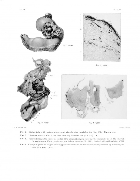
Fig. 4. Syncytium covering a typical villus (No. 808). Stained with hematoxylin and aurantia. The blood corpuscles fill the spaces of the syncytium and fragments of corpuscles lie within the protoplasm of the cells. There are all gradations between complete blood corpuscles and granular protoplasm which take on the same color. X 300.
File history
Click on a date/time to view the file as it appeared at that time.
| Date/Time | Thumbnail | Dimensions | User | Comment | |
|---|---|---|---|---|---|
| current | 17:41, 29 April 2014 | | 2,397 × 3,109 (504 KB) | Z8600021 (talk | contribs) | {{Mall1915 figures}} |
You cannot overwrite this file.
File usage
The following page uses this file: