File:Mall1908a fig80a.jpg
From Embryology
Size of this preview: 568 × 600 pixels. Other resolution: 785 × 829 pixels.
Original file (785 × 829 pixels, file size: 83 KB, MIME type: image/jpeg)
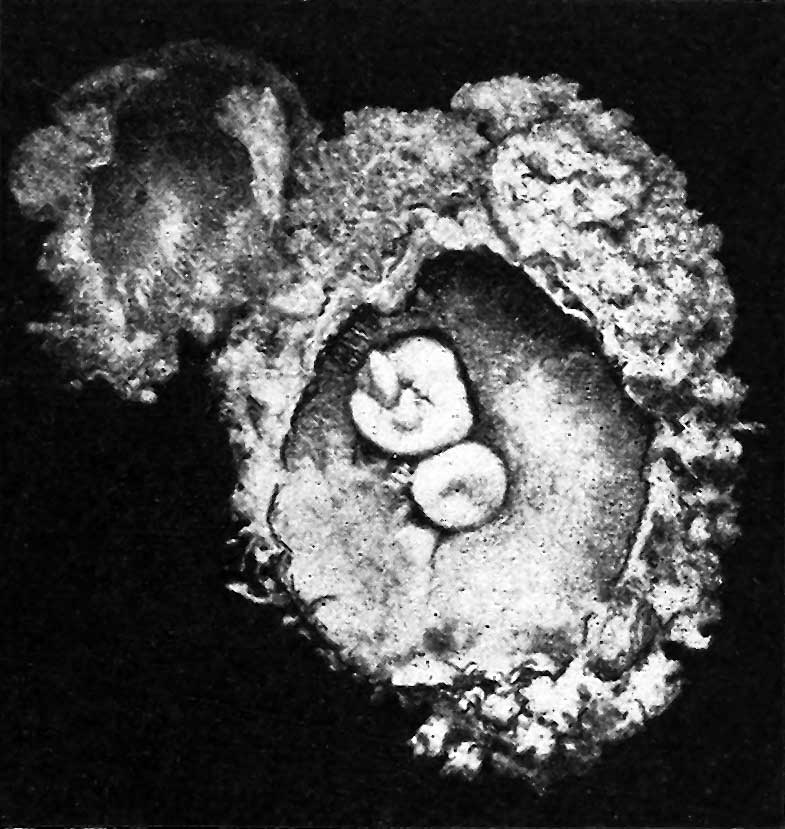
Fig. 80a. Photograph of the ovum and embryo
Enlarged twice. The small additional mass forms a diverticulum of the ovum, the cavity of which communicates with the exoccelom through a narrow opening. The tail of the embryo is twisted.
File history
Click on a date/time to view the file as it appeared at that time.
| Date/Time | Thumbnail | Dimensions | User | Comment | |
|---|---|---|---|---|---|
| current | 12:46, 29 July 2018 | | 785 × 829 (83 KB) | Z8600021 (talk | contribs) | |
| 12:44, 29 July 2018 | 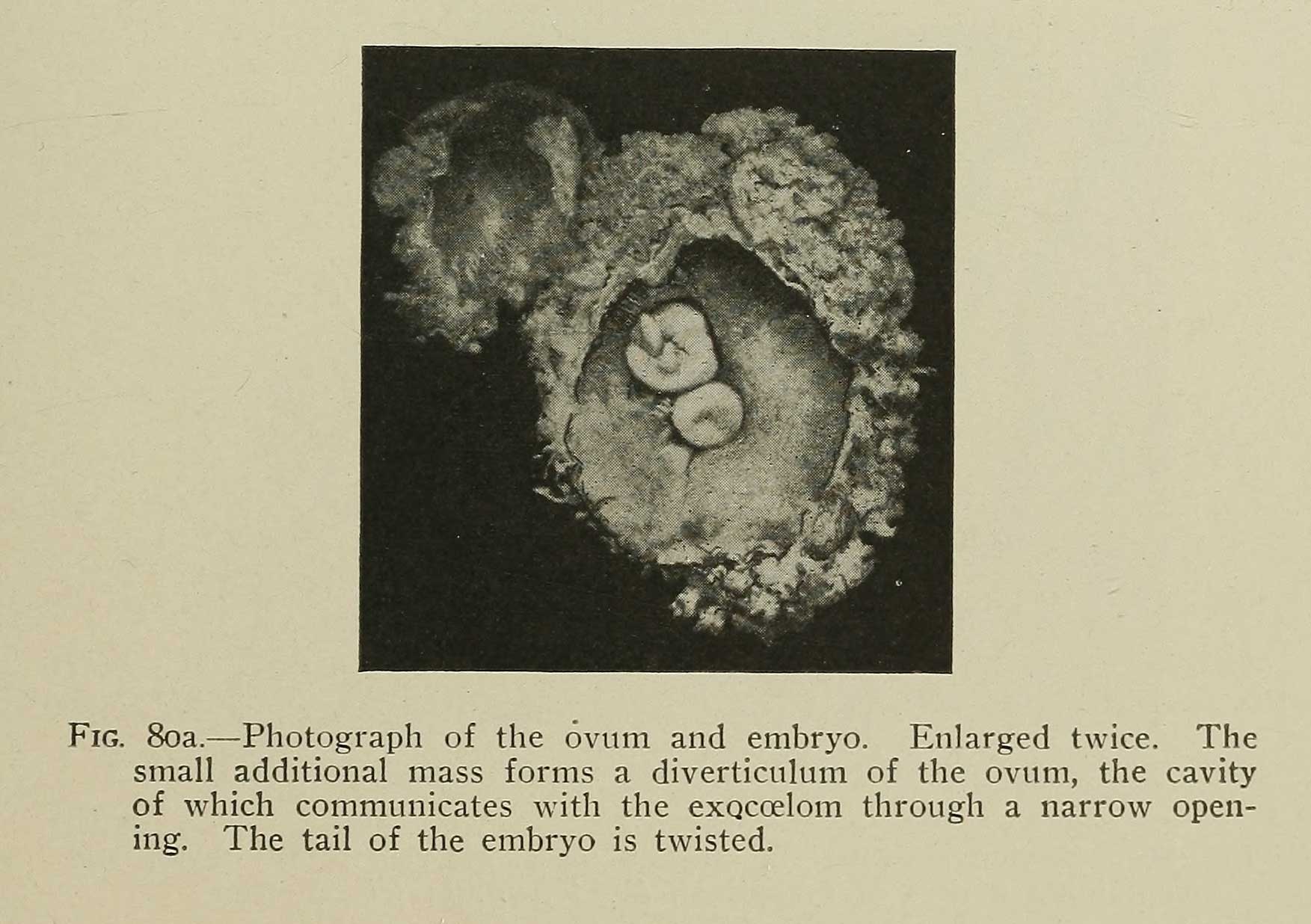 | 1,753 × 1,236 (166 KB) | Z8600021 (talk | contribs) | ==Fig. 80a. Photograph of the ovum and embryo== Enlarged twice. The small additional mass forms a diverticulum of the ovum, the cavity of which communicates with the exoccelom through a narrow opening. The tail of the embryo is twisted. |
You cannot overwrite this file.
File usage
The following page uses this file: