File:Macklin1914 plate04.jpg

Original file (2,435 × 2,640 pixels, file size: 810 KB, MIME type: image/jpeg)
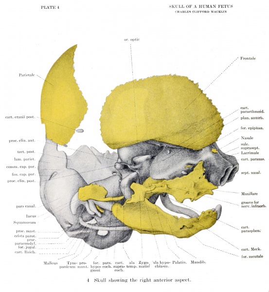
Plate 4. Skull of a 40mm human fetus showing the right anterior aspect
Skull showing the right anterior aspect
Looking at the skull from a more anterior position we see, above, the cavity of the orbit, roofed over by the cartilage of the orbital wing of the sphenoid, and by the orbital portion of the frontal bone (fig. 4) ; limited medially by the shelving posterior portion of the ectethmoid; but widely open downwards and outwards, except where cut off by the zygomatic bone and the maxilla, and but imperfectly closed behind. In the posterior portion of the orbit is seen the elongated optic foramen, and the closeness of apposition of the sphenoidal and ectethmoidal cartilages is to be observed. An open space, communicating freely above with the orbit, below with the cavity of the mouth and medially with that of the pharynx, is seen in front of the ala temporalis, the medial wall of the space being indicated by the imperfectly developed vertical plate of the palatine bone and by the rudimentary medial pterygoid plate. The position of the as yet unformed pterygomaxillary fossa is indicated in this space by the sphenopalatine ganglion (fig. 14) (not shown in the model), and lateral to this, bounded by the incomplete zygomatic arch, are the temporal and zygomatic fossae. One is struck with the lack of prominence of the zygomatic region when compared with the osseous skull, the zygomatic bone and arch being completely overhung by the lateral part of the cranial floor. That this disproportion is partly due to the shallowness of the temporal and zygomatic fossae is evident from a comparison of the model with the bony skull. It would appear that the lateral growth of the ala temporalis of the sphenoid and of the zygomatic process of the maxilla, combined with thickening of the temporalis muscle, are the principal factors which bring about the widening of the temporal and zygomatic fossae, and consequent outpushing of the lateral parts of this area. It is evident from a comparison of the skulls of the newborn and the adult that this change continues till some time after birth.
| Historic Disclaimer - information about historic embryology pages |
|---|
| Pages where the terms "Historic" (textbooks, papers, people, recommendations) appear on this site, and sections within pages where this disclaimer appears, indicate that the content and scientific understanding are specific to the time of publication. This means that while some scientific descriptions are still accurate, the terminology and interpretation of the developmental mechanisms reflect the understanding at the time of original publication and those of the preceding periods, these terms, interpretations and recommendations may not reflect our current scientific understanding. (More? Embryology History | Historic Embryology Papers) |
- Links: plate 1 | plate 2 | plate 3 | plate 4 | plate 5 | plate 6 | fig 6 | fig 7 | plate 7 | fig 8 | fig 9 | plate 8 | fig 10 | plate 9 | fig 11 | fig 12 | plate 10 | plate 11 | fig 14 | Macklin 1914 part 1 | Macklin 1914 part 2 | Skull Development
Reference
Macklin CC. The skull of a human fetus of 40 mm 1. (1914) Amer. J Anat. 16(3): 317-386.
Cite this page: Hill, M.A. (2024, April 27) Embryology Macklin1914 plate04.jpg. Retrieved from https://embryology.med.unsw.edu.au/embryology/index.php/File:Macklin1914_plate04.jpg
- © Dr Mark Hill 2024, UNSW Embryology ISBN: 978 0 7334 2609 4 - UNSW CRICOS Provider Code No. 00098G
File history
Click on a date/time to view the file as it appeared at that time.
| Date/Time | Thumbnail | Dimensions | User | Comment | |
|---|---|---|---|---|---|
| current | 22:33, 20 June 2016 |  | 2,435 × 2,640 (810 KB) | Z8600021 (talk | contribs) |
You cannot overwrite this file.
File usage
The following 2 pages use this file:
