File:Macklin1914 plate01.jpg

Original file (2,184 × 2,602 pixels, file size: 673 KB, MIME type: image/jpeg)
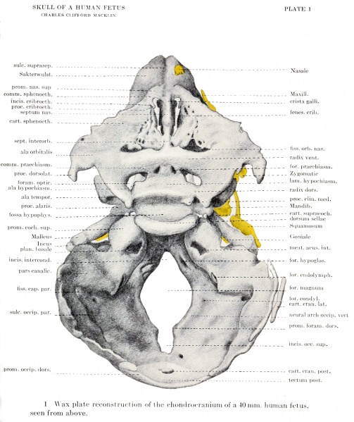
Plate 1. Wax plate reconstruction of the chondrocranium of a 40mm human fetus
Seen from above.
Viewed from above (fig. 1) we note the entire absence of the roof and the extremely rudimentary character of the sides of the cranial vault. The eye meets with an irregular surface of varying depth, surrounded by a broken, ovoid contour, the smaller end being ventral. This surface we recognize as the floor of the primitive brain-case. Its dorsal half is made up of the future posterior cranial fossa, a deep, bowl-like enclosure, the steep sides of which slope rapidly down to an elongated opening in the floor - the primitive foramen magnum. In the ventral wall of this fossa is a trough-like space behind the basilar plate, flanked by two rounded eminences, the partes cochleares of the otic capsule, and terminated above by the horned, ridge-like dorsum sellae, which forms a conspicuous object in the floor of the cranium. Passing forward over this ridge a sideless pit, the hypophyseal fossa, comes into view, which marks the center of the middle cranial fossa, and here, in the region of the body of the sphenoid, the cartilaginous floor is very narrow. Lateral to the corpus sphenoidale is a large, triangular gap in the floor and sidewall of the brain-box, the apex of which meets the side of the sella, while the ventral and dorsal borders are formed by the dorsal border of the ala orbitalis and the cranio-ventral surface of the otic capsule respectively. Forming a lateral, knobbed projection beneath the ala orbitalis is the relatively small ala temporalis, and this is observed to lie ventro-caudal to the plane of the above-mentioned triangular space, just as, in the osseous skull, the greater wing lies below and in front of the plane joining the lesser wing with the petrous portion of the temporal bone. As will be seen later, this plane corresponds in a general way to the situation of a primitive floor and side-wall of this region of the skull, as found in the lizards. Two of the bones which will later wall in this space, viz., the parietal and the squamo-temporaUs, are as yet very rudimentary, while the third, or ala temporalis, has only just commenced to ossify.
| Historic Disclaimer - information about historic embryology pages |
|---|
| Pages where the terms "Historic" (textbooks, papers, people, recommendations) appear on this site, and sections within pages where this disclaimer appears, indicate that the content and scientific understanding are specific to the time of publication. This means that while some scientific descriptions are still accurate, the terminology and interpretation of the developmental mechanisms reflect the understanding at the time of original publication and those of the preceding periods, these terms, interpretations and recommendations may not reflect our current scientific understanding. (More? Embryology History | Historic Embryology Papers) |
- Links: plate 1 | plate 2 | plate 3 | plate 4 | plate 5 | plate 6 | fig 6 | fig 7 | plate 7 | fig 8 | fig 9 | plate 8 | fig 10 | plate 9 | fig 11 | fig 12 | plate 10 | plate 11 | fig 14 | Macklin 1914 part 1 | Macklin 1914 part 2 | Skull Development
Reference
Macklin CC. The skull of a human fetus of 40 mm 1. (1914) Amer. J Anat. 16(3): 317-386.
Cite this page: Hill, M.A. (2024, April 27) Embryology Macklin1914 plate01.jpg. Retrieved from https://embryology.med.unsw.edu.au/embryology/index.php/File:Macklin1914_plate01.jpg
- © Dr Mark Hill 2024, UNSW Embryology ISBN: 978 0 7334 2609 4 - UNSW CRICOS Provider Code No. 00098G
File history
Click on a date/time to view the file as it appeared at that time.
| Date/Time | Thumbnail | Dimensions | User | Comment | |
|---|---|---|---|---|---|
| current | 21:33, 20 June 2016 |  | 2,184 × 2,602 (673 KB) | Z8600021 (talk | contribs) | ==Plate 1. == Wax plato reconstruction of the chondrocranium of a lOniiu. Iiiiinaii fctiLs, - seen from above. ===Reference=== {{Ref-Macklin1914a}} {{Footer}} |
You cannot overwrite this file.
File usage
The following 2 pages use this file:
