File:Macklin1914 fig18.jpg
Original file (1,651 × 1,113 pixels, file size: 284 KB, MIME type: image/jpeg)
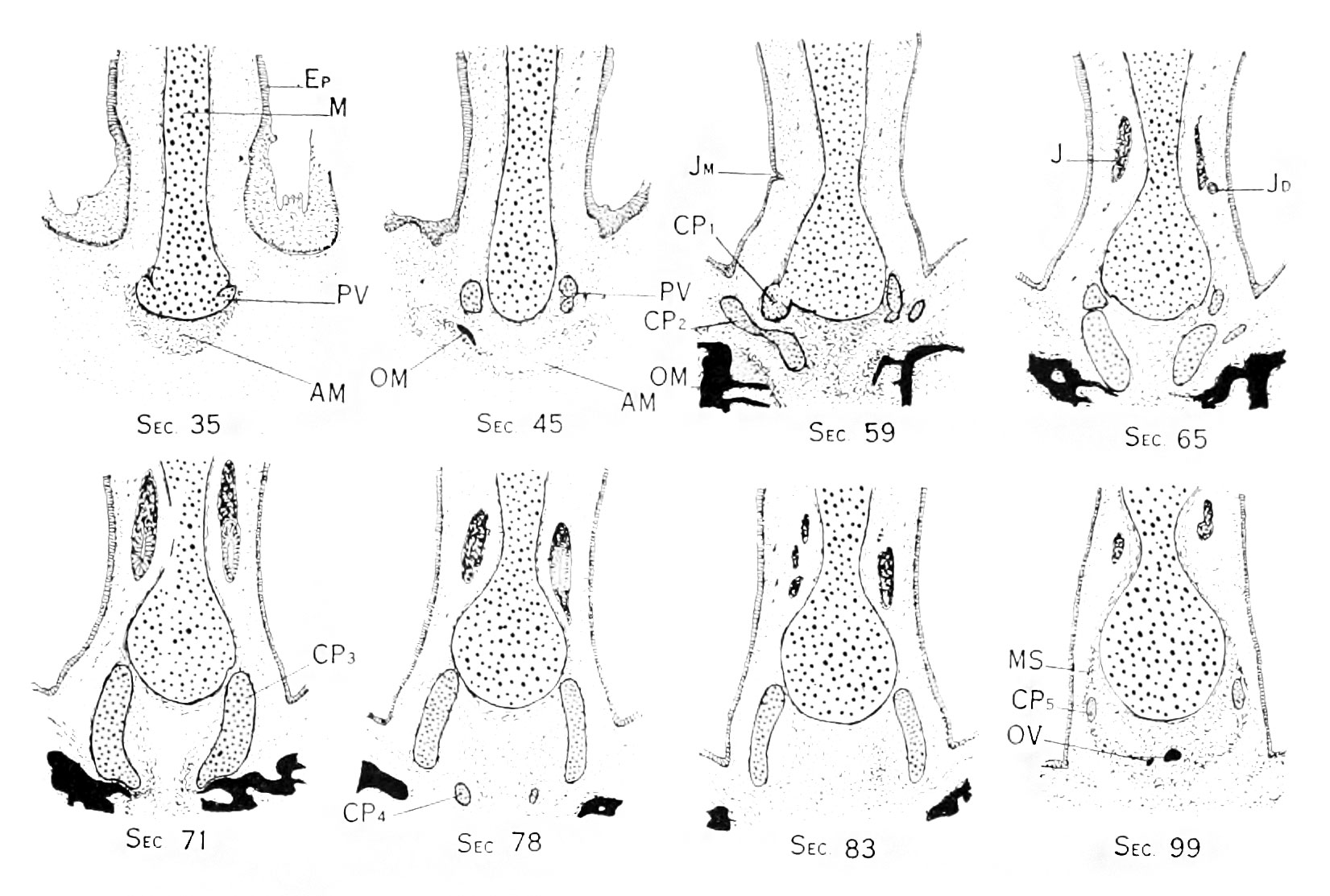
Fig. 18. Is a Series of eight coronal cross-sections through caudal portion of mesethmoid
in the region of the anterior paraseptal cartilages. Sections numbers 35. 45, 59, (35, 71, 78, 83, and 90. Magnif.
Section 35 shows the ventral end of the ventro-lateral process, PV, and its connection with the cauchil border of the mesethmoid. In section 45 the dorsal ends of these processes are shown in cross section, with the ventral extremity of the maxilla OM; the membranous anlage of this bone appears in sections 35 and 45 - AM. Part of the epithelium of the nasal cavity is represented in all the sections Ep. Section 59 is through the ventral end of Jacobson's cartilage and shows the cranio-ventral process of the medial lamina CP1 on the right side attached to the mesethmoid, while the outer lamina, CP2 lies free in the mesenchyme. Owing to the section having been cut somewhat more deeply on the right than on the left side, the left side of the illustration shows a somewhat more dorsal plane than does the right. The meatus of the right Jacobsonian duct opens at Jm in section 59. Section 65 shows, on the right side the dorsal tip of the left outer lamina of Jacobson's cartilage, and medial to this the main part of the inner lamina below, and the cranio-ventral process above. The organ of Jacobson ajipears in this and following sections, and it will be observed that the mesethmoid has become quite thin in its vicinity, ihv. organ lying in a concavity of the cartilage. In section 71 the main lamina of Jacobson's cartilage is seen CP3, and it will be noted that at its caudal extremity the relationship to the maxilla is very intimate. In the next section, No. 78, the caudal process is seen separated from the main mass of the cartilage, and in the next section. No. 83, this process has disappeared. In No. 99 the dorsal tips of the Jacobsonian cartilages are seen CP5, and below the mesethmoid, lying in a mass of condensed mcsenchj'me. the anterior tips of the vomer are seen as two small spicules of bone.
| Historic Disclaimer - information about historic embryology pages |
|---|
 |
- Links: plate 1 | plate 2 | plate 3 | plate 4 | plate 5 | plate 6 | fig 6 | fig 7 | plate 7 | fig 8 | fig 9 | plate 8 | fig 10 | plate 9 | fig 11 | fig 12 | plate 10 | plate 11 | fig 14 | Macklin 1914 part 1 | Macklin 1914 part 2 | Skull Development
Reference
Macklin CC. The skull of a human fetus of 40 mm 1. (1914) Amer. J Anat. 16(3): 317-386.
Cite this page: Hill, M.A. (2024, April 27) Embryology Macklin1914 fig18.jpg. Retrieved from https://embryology.med.unsw.edu.au/embryology/index.php/File:Macklin1914_fig18.jpg
- © Dr Mark Hill 2024, UNSW Embryology ISBN: 978 0 7334 2609 4 - UNSW CRICOS Provider Code No. 00098G
File history
Click on a date/time to view the file as it appeared at that time.
| Date/Time | Thumbnail | Dimensions | User | Comment | |
|---|---|---|---|---|---|
| current | 21:14, 26 June 2016 | | 1,651 × 1,113 (284 KB) | Z8600021 (talk | contribs) |
You cannot overwrite this file.
File usage
The following page uses this file: