File:Macaque Xi at interphase 01.jpg

Original file (1,200 × 615 pixels, file size: 157 KB, MIME type: image/jpeg)
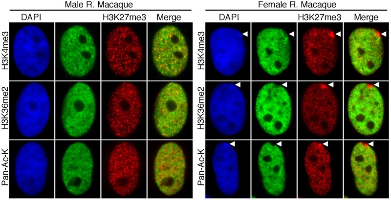
Distribution of euchromatic chromatin marks relative to the macaque Xi at interphase
Typical examples of male and female rhesus macaque (R. Macaque) interphase nuclei showing the distribution of H3K27me3 (red), H3K4me3 (green), H3K36me2 (green) and acetylated lysine (Pan-Ac-K, green) as determined by indirect immunofluorescence.
Nuclei are counterstained with DAPI (blue). White arrowheads in the female images indicate the location of the Xi.
Original image name: Figure 6. Gb-2011-12-4-r37-6-l-new.jpg http://genomebiology.com/2011/12/4/R37/figure/F6
Reference
McLaughlin CR, Chadwick BP. Characterization of DXZ4 conservation in primates implies important functional roles for CTCF binding, array expression and tandem repeat organization on the X chromosome. Genome Biol. 2011 Apr 13;12(4):R37. PMID: 21489251 | Genome Biol.
© 2011 McLaughlin and Chadwick; licensee BioMed Central Ltd. This is an open access article distributed under the terms of the Creative Commons Attribution License (http://creativecommons.org/licenses/by/2.0), which permits unrestricted use, distribution, and reproduction in any medium, provided the original work is properly cited.
File history
Click on a date/time to view the file as it appeared at that time.
| Date/Time | Thumbnail | Dimensions | User | Comment | |
|---|---|---|---|---|---|
| current | 09:53, 29 May 2011 |  | 1,200 × 615 (157 KB) | S8600021 (talk | contribs) | ==Distribution of euchromatic chromatin marks relative to the macaque Xi at interphase== Typical examples of male and female rhesus macaque (R. Macaque) interphase nuclei showing the distribution of H3K27me3 (red), H3K4me3 (green), H3K36me2 (green) and a |
You cannot overwrite this file.
File usage
There are no pages that use this file.