File:MERS-CoV EM1.jpg: Difference between revisions
From Embryology
| Line 8: | Line 8: | ||
{{Footer}} | {{Footer}} | ||
[[Category:Virus]][[Category: | [[Category:Virus]][[Category:Electron Microscopy]] | ||
Revision as of 00:38, 21 January 2020
MERS-CoV particles as seen by negative stain electron microscopy, coronavirus virions contain characteristic "corona" of club-like projections from the viral membrane.
Reference
CNational Center for Immunization and Respiratory Diseases (NCIRD), Division of Viral Diseases
CDC Source: Cynthia Goldsmith/Maureen Metcalfe/Azaibi Tamin
Cite this page: Hill, M.A. (2024, April 27) Embryology MERS-CoV EM1.jpg. Retrieved from https://embryology.med.unsw.edu.au/embryology/index.php/File:MERS-CoV_EM1.jpg
- © Dr Mark Hill 2024, UNSW Embryology ISBN: 978 0 7334 2609 4 - UNSW CRICOS Provider Code No. 00098G
File history
Click on a date/time to view the file as it appeared at that time.
| Date/Time | Thumbnail | Dimensions | User | Comment | |
|---|---|---|---|---|---|
| current | 00:31, 21 January 2020 | 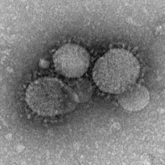 | 537 × 537 (68 KB) | Z8600021 (talk | contribs) | MERS-CoV particles as seen by negative stain electron microscopy. Virions contain characteristic club-like projections emanating from the viral membrane. ===Reference=== CDC Source: Cynthia Goldsmith/Maureen Metcalfe/Azaibi Tamin {{Footer}} Category:VirusCategory:EM |
You cannot overwrite this file.
File usage
The following 3 pages use this file: