File:Lymph node structure 02.jpg: Difference between revisions
From Embryology
No edit summary |
m (→Reference) |
||
| (7 intermediate revisions by the same user not shown) | |||
| Line 1: | Line 1: | ||
==Lymph | ==Lymph Node Structure== | ||
Schematic representation of the organization of a lymph node | Schematic representation of the organization of a lymph node. | ||
* '''Afferent lymphatics''' enter lymph nodes and deliver lymph to the '''subcapsular sinus''' (SCS), which forms a channel around the periphery of the lymph node. | |||
* '''Lymphatic sinuses''' run from the SCS through the cortex to the medulla | |||
* '''Efferent lymphatics''' exit the lymph node via efferent lymphatic vessels on the opposite (hilar) side of the organ. | |||
* '''High endothelial venules''' (HEVs) - in the paracortex and cortical ridge, specialized vessels allow entry of leukocytes from the blood. | |||
* Blood vessels enter and exit the lymph node on the hilar side, and snake through the lymph node like the branches of a tree. | |||
* '''B cell follicles''' containing follicular dendritic cell (FDC) networks are arranged in the lymph node cortex and are separated from the SCS by a layer of marginal reticular cells (MRC). | |||
* '''T cells zones''' in the paracortex, which contain many fibroblastic reticular cells (FRC), are separated by the cortical ridge, an area rich in T cells, dendritic cells (DCs), blood vessels, and FRC. | |||
Scale bars represent 200 μM. | Scale bars represent 200 μM. | ||
{{Lymph node cartoons}} | |||
===Reference=== | ===Reference=== | ||
{{#pmid:19644499}} | |||
[http://www.microbiol.unimelb.edu.au/research/immunology/s_mueller.html Mueller] | [http://www.microbiol.unimelb.edu.au/research/immunology/s_mueller.html Mueller] | ||
====Copyright==== | |||
[[File_talk:Lymph_node_structure_02.jpg|Permissions]] | [[File_talk:Lymph_node_structure_02.jpg|Permissions]] | ||
{{Footer}} | |||
[[Category:Immune]] | [[Category:Immune]] | ||
Latest revision as of 18:39, 30 April 2018
Lymph Node Structure
Schematic representation of the organization of a lymph node.
- Afferent lymphatics enter lymph nodes and deliver lymph to the subcapsular sinus (SCS), which forms a channel around the periphery of the lymph node.
- Lymphatic sinuses run from the SCS through the cortex to the medulla
- Efferent lymphatics exit the lymph node via efferent lymphatic vessels on the opposite (hilar) side of the organ.
- High endothelial venules (HEVs) - in the paracortex and cortical ridge, specialized vessels allow entry of leukocytes from the blood.
- Blood vessels enter and exit the lymph node on the hilar side, and snake through the lymph node like the branches of a tree.
- B cell follicles containing follicular dendritic cell (FDC) networks are arranged in the lymph node cortex and are separated from the SCS by a layer of marginal reticular cells (MRC).
- T cells zones in the paracortex, which contain many fibroblastic reticular cells (FRC), are separated by the cortical ridge, an area rich in T cells, dendritic cells (DCs), blood vessels, and FRC.
Scale bars represent 200 μM.
- Lymph Node Cartoons: Detailed structure | Cartoon with Histology | Lymphocyte traffic | Simple structure | Simple node anatomy | Wiki node image | Internal structure | Mesenteric lymph node | Histology | Gallery | Lymph Node Development
Reference
Mueller SN & Germain RN. (2009). Stromal cell contributions to the homeostasis and functionality of the immune system. Nat. Rev. Immunol. , 9, 618-29. PMID: 19644499 DOI. Mueller
Copyright
Cite this page: Hill, M.A. (2024, May 20) Embryology Lymph node structure 02.jpg. Retrieved from https://embryology.med.unsw.edu.au/embryology/index.php/File:Lymph_node_structure_02.jpg
- © Dr Mark Hill 2024, UNSW Embryology ISBN: 978 0 7334 2609 4 - UNSW CRICOS Provider Code No. 00098G
File history
Click on a date/time to view the file as it appeared at that time.
| Date/Time | Thumbnail | Dimensions | User | Comment | |
|---|---|---|---|---|---|
| current | 18:53, 22 February 2012 | 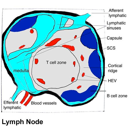 | 460 × 463 (54 KB) | Z8600021 (talk | contribs) | ==Lymph node structure== Schematic representation of the organization of a lymph node (left panel). Afferent lymphatics enter lymph nodes and deliver lymph to the subcapsular sinus (SCS), which forms a channel around the periphery of the lymph node. Lymp |
You cannot overwrite this file.
File usage
The following 4 pages use this file: