File:Lymph node histology01.jpg
From Embryology
Size of this preview: 705 × 599 pixels.
Original file (800 × 680 pixels, file size: 282 KB, MIME type: image/jpeg)
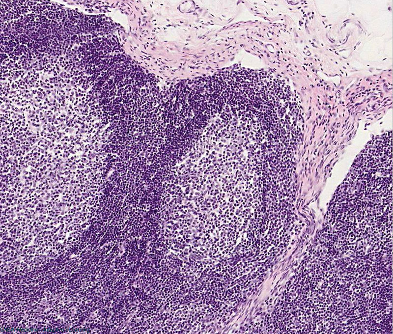
Lymph Node Histology
- Capsule - dense connective tissue.
- Trabeculae - connective tissue extending from capsule.
- Paratrabecular sinus - lymph-filled space surrounding trabeculae.
- Subcapsular sinus - afferent lymphatic vessels empty into this space (continuous with medullary sinus).
- Germinal centre - pale spherical region, located at cortex.
- Mantle zone - dark spherical region surrounding germinal centre.
Source
Department of Anatomy teaching set in UNSW Virtual Slide Repository.
File history
Click on a date/time to view the file as it appeared at that time.
| Date/Time | Thumbnail | Dimensions | User | Comment | |
|---|---|---|---|---|---|
| current | 08:41, 28 February 2011 | | 800 × 680 (282 KB) | S8600021 (talk | contribs) | ==Lymph Node Histology== Department of Anatomy teaching set in UNSW Virtual Slide Repository. Category:Histology Category:Immune |
You cannot overwrite this file.
File usage
There are no pages that use this file.