File:Lymph node cartoon.jpg
Lymph_node_cartoon.jpg (600 × 450 pixels, file size: 66 KB, MIME type: image/jpeg)
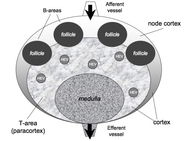
Schematic representation of a lymph node internal architecture
Human lymph nodes are bean-shaped structures that range in size from a few millimeters to about 1-2 cm in their normal state. Internally, two main regions can be distinguished: the medulla and the cortex. The cortex can be further divided into an inner part, the paracortex (also called the T cell area), rich in T lymphocytes and an outer area, the node cortex that includes the B cell area consisting of follicles and germinal centers, where B cells are activated and differentiate. T and B areas are identified by high concentrations of different chemokines (CCR7 and CXCR5, respectively) secreted by local stromal cells. The whole structure is supported by a dense network of fibroblastic reticular cells that encloses small lymphatic channels of 10-15 μm in diameter along which small molecules are thought to diffuse.
- T cell area - the paracortex
- B cell area the node cortex
- HEV - high endothelial venules are located inside the paracortex
A number of follicles are located in the node cortex, whereas the T cell area occupies the inner part.
The medulla consists of a part of the T-area that allows cells to exit by the efferent vessel. T and B cells enter the lymph node mainly from the blood, through HEV located inside the paracortex.
Absence of Infection
In the absence of an antigenic challenge, T and B cells randomly scan their respective areas for ~24-48 h before exiting the lymph node. Entrance of antigens into the lymph node triggers a series of events leading to antigen recognition and the activation of an immune response.
Infection
- During an infection, lymphocyte recruitment from the periphery is enhanced due to a widening of the primary arteriole feeding the lymph node.
- Once inside the lymph node, B and T cells rapidly home in their own compartments, following a specific chemotactic gradient.
- T helper cells (TH) are the fastest (average velocity of 11 μm/min)
- B cells (average velocity of 6 μm/min)
- Dendritic cells (DCs) (average velocity of 3 μm/min)
(Above text modified from original article)
- Lymph Node Cartoons: Detailed structure | Cartoon with Histology | Lymphocyte traffic | Simple structure | Simple node anatomy | Wiki node image | Internal structure | Mesenteric lymph node | Histology | Gallery | Lymph Node Development
Reference
<pubmed>19939270</pubmed>| BMC Bioinformatics.
Baldazzi et al. BMC Bioinformatics 2009 10:387 doi:10.1186/1471-2105-10-387
© 2009 Baldazzi et al; licensee BioMed Central Ltd.
This is an Open Access article distributed under the terms of the Creative Commons Attribution License (http://creativecommons.org/licenses/by/2.0), which permits unrestricted use, distribution, and reproduction in any medium, provided the original work is properly cited.
Original file name: 1471-2105-10-387-3.jpg
File history
Click on a date/time to view the file as it appeared at that time.
| Date/Time | Thumbnail | Dimensions | User | Comment | |
|---|---|---|---|---|---|
| current | 13:05, 22 December 2010 | | 600 × 450 (66 KB) | S8600021 (talk | contribs) | ==Schematic representation of a lymph node internal architecture== * T cell area - the paracortex * B cell area the node cortex A number of follicles are located in the node cortex, whereas the T cell area occupies the inner part. The medulla consist |
You cannot overwrite this file.
File usage
The following 4 pages use this file: