File:Lockwood1887b fig32.jpg
From Embryology
No higher resolution available.
Lockwood1887b_fig32.jpg (500 × 329 pixels, file size: 55 KB, MIME type: image/jpeg)
Plate II
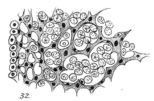
Fig. 32. Genital mass of rabbit, commencement of fourteenth day, to show stroma of branched anastomosing cells, and large, pale, granular cells in its meshes, 7 Hartnack. 4 Eye p.
Reference
Lockwood CB. Development and transition of the testis, normal and abnormal. (1887) J Anat. 22(1): 38-77. PMID 17231729
Cite this page: Hill, M.A. (2024, April 27) Embryology Lockwood1887b fig32.jpg. Retrieved from https://embryology.med.unsw.edu.au/embryology/index.php/File:Lockwood1887b_fig32.jpg
- © Dr Mark Hill 2024, UNSW Embryology ISBN: 978 0 7334 2609 4 - UNSW CRICOS Provider Code No. 00098G
File history
Click on a date/time to view the file as it appeared at that time.
| Date/Time | Thumbnail | Dimensions | User | Comment | |
|---|---|---|---|---|---|
| current | 19:50, 14 April 2020 | | 500 × 329 (55 KB) | Z8600021 (talk | contribs) |
You cannot overwrite this file.
File usage
The following page uses this file: