File:Liver sinusoidal endothelial cell fenestrations.jpg

Original file (926 × 474 pixels, file size: 184 KB, MIME type: image/jpeg)
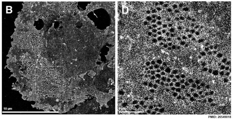
Liver Sinusoidal Endothelial Cell Fenestrations
Liver sinusoidal endothelial cells (LSECs) act as a filter between blood and the hepatocytes. LSECs are highly fenestrated cells; they contain transcellular pores with diameters between 50 to 200 nm. The small sizes of the fenestrae have so far prohibited any functional analysis with standard and advanced light microscopy techniques. Organised as “sieve plates”, assemblies of fenestrations in regions where LSECs are extremely thin (approx. 300 nm on average).
(B,D) dSTORM images of a fixed rat LSECs stained with Vybrant DiD. The cells were mounted in the reducing buffer OSS + MEA. (D) show enlarged view of the corresponding ROIs shown in (B). The exposure time for a single dSTORM frame was 20 ms and 15000 frames were processed to reconstruct the images shown in (B,D).
Reference
Mönkemöller V, Øie C, Hübner W, Huser T & McCourt P. (2015). Multimodal super-resolution optical microscopy visualizes the close connection between membrane and the cytoskeleton in liver sinusoidal endothelial cell fenestrations. Sci Rep , 5, 16279. PMID: 26549018 DOI.
Mönkemöller, V. et al. Multimodal super-resolution optical microscopy visualizes the close connection between membrane and the cytoskeleton in liver sinusoidal endothelial cell fenestrations. Sci. Rep. 5, 16279; doi: 10.1038/srep16279 (2015).
Copyright
This work is licensed under a Creative Commons Attribution 4.0 International License. The images or other third party material in this article are included in the article’s Creative Commons license, unless indicated otherwise in the credit line; if the material is not included under the Creative Commons license, users will need to obtain permission from the license holder to reproduce the material. To view a copy of this license, visit http://creativecommons.org/licenses/by/4.0/
Srep16279-f2.jpg
Cite this page: Hill, M.A. (2024, April 27) Embryology Liver sinusoidal endothelial cell fenestrations.jpg. Retrieved from https://embryology.med.unsw.edu.au/embryology/index.php/File:Liver_sinusoidal_endothelial_cell_fenestrations.jpg
- © Dr Mark Hill 2024, UNSW Embryology ISBN: 978 0 7334 2609 4 - UNSW CRICOS Provider Code No. 00098G
File history
Click on a date/time to view the file as it appeared at that time.
| Date/Time | Thumbnail | Dimensions | User | Comment | |
|---|---|---|---|---|---|
| current | 22:25, 14 March 2016 | | 926 × 474 (184 KB) | Z8600021 (talk | contribs) | |
| 22:24, 14 March 2016 |  | 926 × 926 (257 KB) | Z8600021 (talk | contribs) |
You cannot overwrite this file.
File usage
The following 2 pages use this file: