File:Liver polyploidy 01.jpg: Difference between revisions
From Embryology
No edit summary |
No edit summary |
||
| Line 1: | Line 1: | ||
==Liver Polyploidy== | ==Liver Polyploidy== | ||
Polyploid hepatocyte mitoses with multipolar spindles and chromosome segregation defects | Polyploid hepatocyte mitoses in vitro with multipolar spindles and chromosome segregation defects. | ||
* '''A''' - bipolar spindles were established and maintained by centrosome clustering (in about half of tetraploid mitoses) | |||
* '''B C''' - centrosomes oriented on 3-4 distinct poles (reflecting either true multipolar spindles or alignment of prometaphase chromosomes from binucleated hepatocytes) | |||
* '''D''' - octaploid hepatocytes established multipolar spindles with as many as 8 poles | |||
* '''E''' - about 1% of cells in anaphase or telophase were oriented with tripolar spindles | |||
* '''F''' - hepatocytes with 2 discrete mitotic spindles synchronized in metaphase | |||
* '''G''' - hepatocytes with 2 discrete mitotic spindles synchronized in anaphase | |||
* '''I - L''' - mitotic figures similar to those seen in cultured hepatocytes were also seen during hepatocyte proliferation in vivo | |||
Original File Name: Fig. 3 Nature09414-f3.2.jpg | Original File Name: Fig. 3 Nature09414-f3.2.jpg | ||
Revision as of 10:26, 14 August 2011
Liver Polyploidy
Polyploid hepatocyte mitoses in vitro with multipolar spindles and chromosome segregation defects.
- A - bipolar spindles were established and maintained by centrosome clustering (in about half of tetraploid mitoses)
- B C - centrosomes oriented on 3-4 distinct poles (reflecting either true multipolar spindles or alignment of prometaphase chromosomes from binucleated hepatocytes)
- D - octaploid hepatocytes established multipolar spindles with as many as 8 poles
- E - about 1% of cells in anaphase or telophase were oriented with tripolar spindles
- F - hepatocytes with 2 discrete mitotic spindles synchronized in metaphase
- G - hepatocytes with 2 discrete mitotic spindles synchronized in anaphase
- I - L - mitotic figures similar to those seen in cultured hepatocytes were also seen during hepatocyte proliferation in vivo
Original File Name: Fig. 3 Nature09414-f3.2.jpg
Reference
<pubmed>20861837</pubmed>| PMC2967727 | Nature
Reprinted by permission from Macmillan Publishers Ltd.
File history
Click on a date/time to view the file as it appeared at that time.
| Date/Time | Thumbnail | Dimensions | User | Comment | |
|---|---|---|---|---|---|
| current | 10:15, 14 August 2011 | 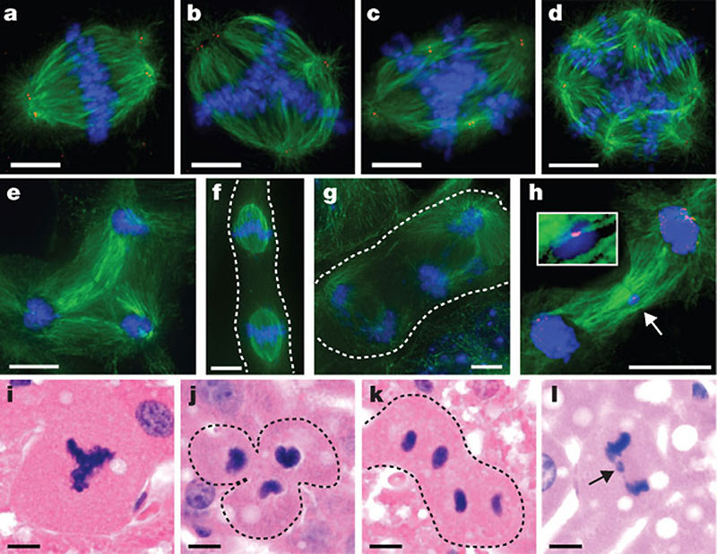 | 800 × 619 (119 KB) | S8600021 (talk | contribs) | ==Liver Polyploidy== Polyploid hepatocyte mitoses with multipolar spindles and chromosome segregation defects Original File Name: Fig. 3 Nature09414-f3.2.jpg http://www.nature.com/authors/policies/license.html |
You cannot overwrite this file.
File usage
The following 2 pages use this file: