File:Liver SEM01.jpg
Original file (2,000 × 1,333 pixels, file size: 350 KB, MIME type: image/jpeg)
Liver Bile Canaliculi and Sinusoids SEM
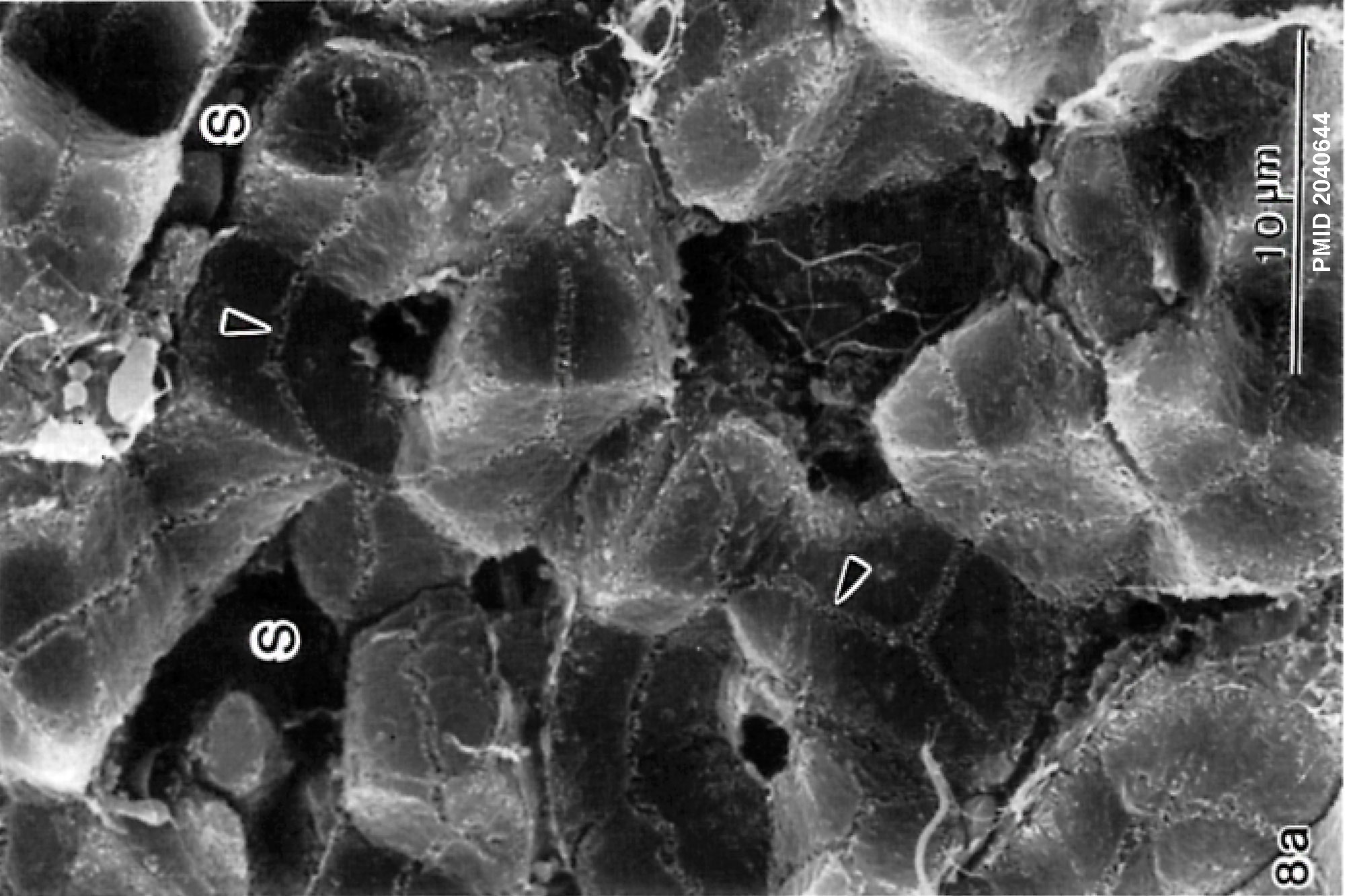
Scanning electron micrographs of a fractured surface of rat livers. (a) Control liver. Note the chicken wire-like anastomosing network of bile canaliculi. Canalicular lumina are uniformly filled with microvilli.
arrowheads - bile canaliculi (channels shown shown as narrow paired lines)
S - sinusoid
- Links:
Reference
Watanabe N, Tsukada N, Smith CR & Phillips MJ. (1991). Motility of bile canaliculi in the living animal: implications for bile flow. J. Cell Biol. , 113, 1069-80. PMID: 2040644
Copyright
Rockefeller University Press - Copyright Policy This article is distributed under the terms of an Attribution–Noncommercial–Share Alike–No Mirror Sites license for the first six months after the publication date (see http://www.jcb.org/misc/terms.shtml). After six months it is available under a Creative Commons License (Attribution–Noncommercial–Share Alike 4.0 Unported license, as described at https://creativecommons.org/licenses/by-nc-sa/4.0/ ). (More? Help:Copyright Tutorial)
Figure 8 panel A cropped, resized and relabelled with PMID.
Cite this page: Hill, M.A. (2024, April 26) Embryology Liver SEM01.jpg. Retrieved from https://embryology.med.unsw.edu.au/embryology/index.php/File:Liver_SEM01.jpg
- © Dr Mark Hill 2024, UNSW Embryology ISBN: 978 0 7334 2609 4 - UNSW CRICOS Provider Code No. 00098G
File history
Click on a date/time to view the file as it appeared at that time.
| Date/Time | Thumbnail | Dimensions | User | Comment | |
|---|---|---|---|---|---|
| current | 15:10, 18 May 2018 | | 2,000 × 1,333 (350 KB) | Z8600021 (talk | contribs) | J Cell Biol. 1991 Jun;113(5):1069-80. Motility of bile canaliculi in the living animal: implications for bile flow. Watanabe N1, Tsukada N, Smith CR, Phillips MJ. Author information Abstract Modern fluorescence microscopic techniques were used to image... |
You cannot overwrite this file.
File usage
The following page uses this file: