File:Lisser1911 fig38.jpg: Difference between revisions
From Embryology
mNo edit summary |
|||
| (One intermediate revision by the same user not shown) | |||
| Line 1: | Line 1: | ||
==Fig. 38 Wax model of laryngeal region in human Embryo 22== | ==Fig. 38 Wax model of laryngeal region in human Embryo 22== | ||
Embryo no. {{CE22}} (20 mm.). (drawn from the left side) | |||
Latest revision as of 10:14, 16 May 2017
Fig. 38 Wax model of laryngeal region in human Embryo 22
Embryo no. 22 (20 mm.). (drawn from the left side)
- Links: fig 1 | fig 2 | fig 3 | fig 4 | fig 5 | fig 6 | fig 7 | fig 8 | fig 9 | fig 10 | fig 11 | fig 12 | fig 13 | fig 14 | Lisser 1911
Reference
Lisser H. Studies on the development of the human larynx. (1911) Amer. J Anat. 12: 27-66.
Cite this page: Hill, M.A. (2024, April 26) Embryology Lisser1911 fig38.jpg. Retrieved from https://embryology.med.unsw.edu.au/embryology/index.php/File:Lisser1911_fig38.jpg
- © Dr Mark Hill 2024, UNSW Embryology ISBN: 978 0 7334 2609 4 - UNSW CRICOS Provider Code No. 00098G
File history
Click on a date/time to view the file as it appeared at that time.
| Date/Time | Thumbnail | Dimensions | User | Comment | |
|---|---|---|---|---|---|
| current | 21:25, 15 June 2016 | 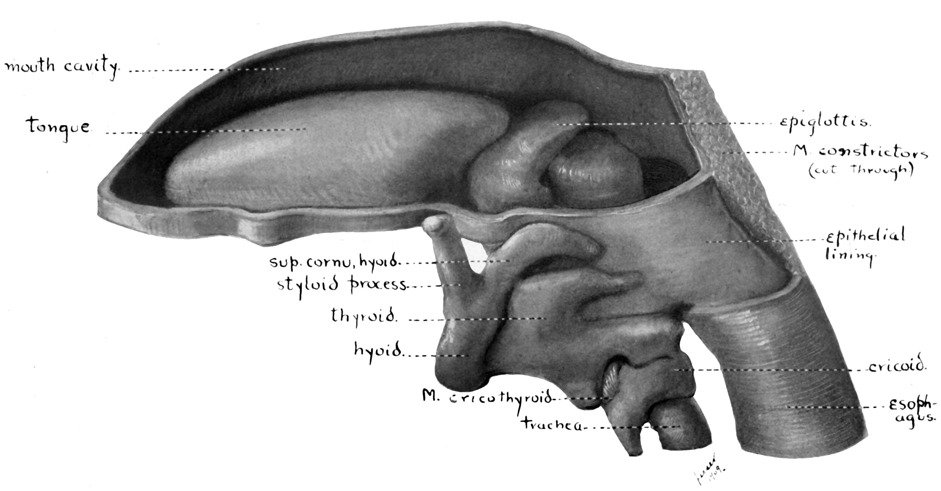 | 3,041 × 1,586 (351 KB) | Z8600021 (talk | contribs) |
You cannot overwrite this file.
File usage
The following 3 pages use this file: