File:Limbal stem cell niche cartoon PMID17211449.jpg
Limbal_stem_cell_niche_cartoon_PMID17211449.jpg (800 × 579 pixels, file size: 52 KB, MIME type: image/jpeg)
Limbal stem cell niche cartoon
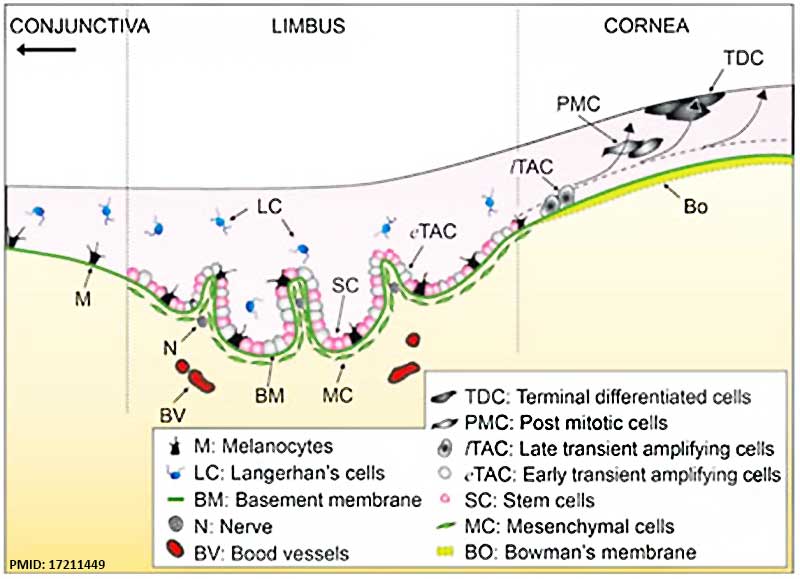
Limbal epithelial stem cells (SC) are located at the limbal basal layer. In this epithelial level, there are several other cell types in the vicinity such as the immediate progeny, i.e., early transient amplifying cells (eTAC), melanocytes (M), and Langerhan's cells (LC). It remains to be determined whether these cell types act as niche cells. It is believed that eTAC will be destined for progeny production by differentiating into late TACs (lTAC) located at the corneal basal layer, then into suprabasal post-mitotic cells (PMC), and finally into superficial terminally differentiated cells (TDC). The limbal basement membrane (BM) separating the epithelium from the underlying stroma has several unique components. The subjacent limbal stroma contains mesenchymal cells (MC), which may also serve as niche cells. Because the limbal stroma is highly innervated and vascularized, the respective role of nerves (N) and blood vessels (BV) in the niche remains to be defined.
- Links: Cornea Development
Reference
<pubmed>17211449</pubmed>| Cell Research
Copyright
Reprinted by permission from Macmillan Publishers Ltd: Cell Research (<pubmed>17211449</pubmed>), copyright (2007)
Thank you for placing your order through Copyright Clearance Center's RightsLink service. Nature Publishing Group has partnered with RightsLink to license its content. This notice is a confirmation that your order was successful.
Your order details and publisher terms and conditions are available by clicking the link below: http://s100.copyright.com/CustomerAdmin/PLF.jsp?ref=a9604785-cf40-4da0-95c8-b48612479610
Order Details Licensee: Mark A Hill License Date: Aug 29, 2014 License Number: 3458520757960 Publication: Cell Research Title: Niche regulation of corneal epithelial stem cells at the limbus Type Of Use: post on a website
Figure 2 http://www.nature.com/cr/journal/v17/n1/fig_tab/7310137f2.html#figure-title Adjusted in size and labelling.
File history
Click on a date/time to view the file as it appeared at that time.
| Date/Time | Thumbnail | Dimensions | User | Comment | |
|---|---|---|---|---|---|
| current | 12:21, 30 August 2014 |  | 800 × 579 (52 KB) | Z8600021 (talk | contribs) | ==Limbal stem cell niche cartoon== Limbal epithelial stem cells (SC) are located at the limbal basal layer. In this epithelial level, there are several other cell types in the vicinity such as the immediate progeny, i.e., early transient amplifying ce... |
You cannot overwrite this file.
File usage
The following page uses this file:
