File:Leydig cell PMID13693345 EM02.jpg
Original file (1,359 × 957 pixels, file size: 341 KB, MIME type: image/jpeg)
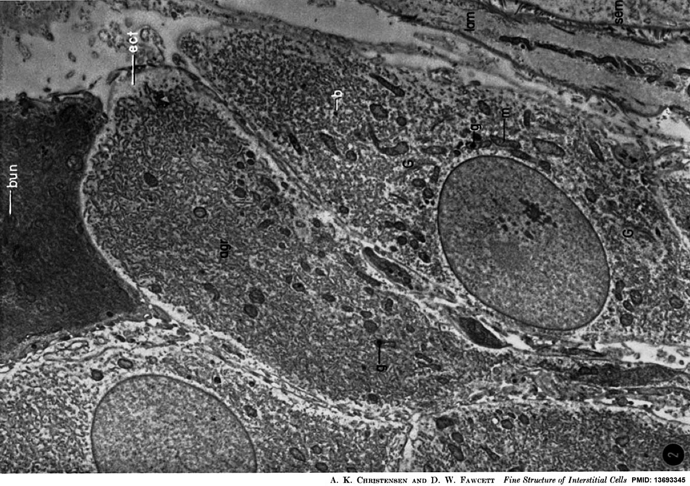
A low-power electron micrograph showing several interstitial cells, one of which (above) contains cytoplasm of considerably greater density than the others. The most striking feature of the interstitial cells is the abundant agranular endoplasmic reticulmn (agr), which fills their cytoplasm and is in the form of a network of interconnected tubules. At the periphery of thc cells is an ectoplasmic zone (ect), which is relatively free of endoplasmic reticulum. The cytoplasm also contains mitochondria (m), in which large, homogeneous granules (g) sometimes occur (see Fig. 10). Various extensions of the Golgi zone (G) arc seen around the nucleus. Small, dark bodies (b) of unknown nature are also found in the cytoplasm. The denser cell contains an oblique section through a bundle (bun) of minute tubules, but it is difficult to make out detail because of the great density of the cytoplasm. The edge of a seminiferous tubule (sere) is seen at lower right, and is flanked by one of the cells (lain) which contribute to the lamina propria of the tubule. X 6,200.
J Biophys Biochem Cytol. 1961 Mar;9:653-70.
The normal fine structure of opossum testicular interstitial cells.
CHRISTENSEN AK, FAWCETT DW.
Abstract
The interstitial tissue of the opossum testis includes interstitial or Leydig cells, macrophages, and small cells which morphologically resemble mesenchymal cells. The latter are thought to give rise to mature interstitial cells. The most prominent feature of the interstitial cell cytoplasm is an exceedingly abundant agranular endoplasmic reticulum. This reticulum is generally in the form of a meshwork of interconnected tubules about 300 to 450 A in diameter, but occasionally it assumes the form of flattened, fenestrated cisternae resembling those of pancreatic acinar cells, except for the lack of ribonucleoprotein particles on the surface of the membranes. The interstitial cells vary considerably in their cytoplasmic density. The majority are quite light, but some appear extremely dense, and in addition usually have a more irregular cell surface, with numerous small pseudopodia. These differences may well reflect variations in physiological state. Cytoplasmic structures previously interpreted as "crystalloids" consist of long bundles of minute parallel tubules, each about 180 A in diameter, which seem to be local differentiations of the endoplasmic reticulum. The mitochondria are rod-shaped, and contain a moderately complex internal membrane structure, and also occasional large inclusions that are spherical and homogeneous. The prominent juxtanuclear Golgi complex contains closely packed flattened sacs and small vesicles. The results of the present study, coupled with biochemical evidence from other laboratories, make it seem highly probable that the agranular endoplasmic reticulum is involved in the synthesis of the steroid hormones produced by the interstitial cell. This finding therefore constitutes one of the first functions of the agranular reticulum for which there is good morphological and biochemical evidence.
PMID 13693345
Copyright
Rockefeller University Press - Copyright Policy This article is distributed under the terms of an Attribution–Noncommercial–Share Alike–No Mirror Sites license for the first six months after the publication date (see http://www.jcb.org/misc/terms.shtml). After six months it is available under a Creative Commons License (Attribution–Noncommercial–Share Alike 4.0 Unported license, as described at https://creativecommons.org/licenses/by-nc-sa/4.0/ ). (More? Help:Copyright Tutorial)
File history
Click on a date/time to view the file as it appeared at that time.
| Date/Time | Thumbnail | Dimensions | User | Comment | |
|---|---|---|---|---|---|
| current | 17:13, 7 August 2014 | | 1,359 × 957 (341 KB) | Z8600021 (talk | contribs) | J Biophys Biochem Cytol. 1961 Mar;9:653-70. The normal fine structure of opossum testicular interstitial cells. CHRISTENSEN AK, FAWCETT DW. Abstract The interstitial tissue of the opossum testis includes interstitial or Leydig cells, macrophages, and s... |
You cannot overwrite this file.
File usage
The following page uses this file: