File:LewisHarrison1966 fig06.jpg
From Embryology
Size of this preview: 289 × 598 pixels. Other resolution: 1,000 × 2,069 pixels.
Original file (1,000 × 2,069 pixels, file size: 325 KB, MIME type: image/jpeg)
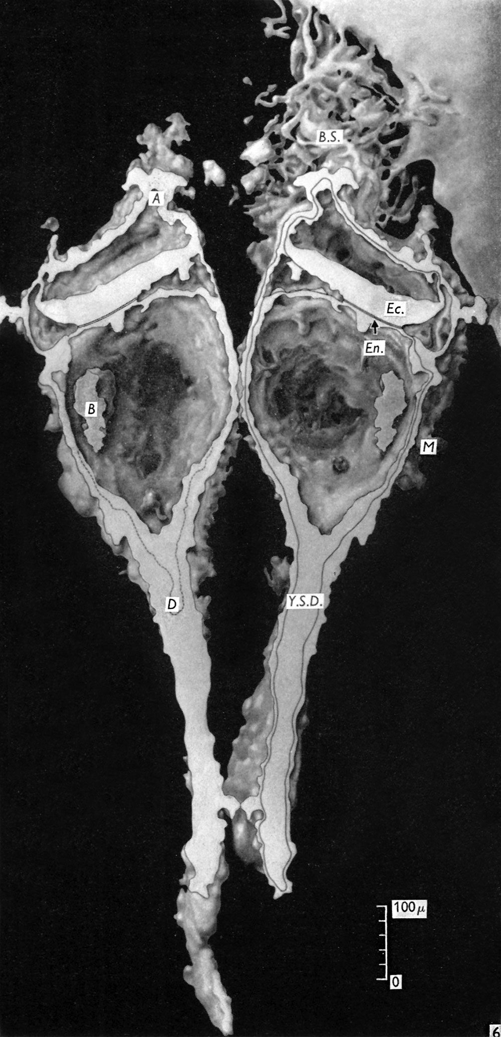
Fig. 6. Diagram of a reconstruction model of the embryo
A, Amniotic duct; B, collection of blood cells in yolk-sac lumen; B.S., connecting stalk; D, diverticulum of yolk-sac passing into yolk-sac duct (Y.S.D.); Ec., Ectoderm; En., endoderm; M, extra-embryonic mesoderm.
Reference
Lewis BV. and Harrison RG. A presomite human embryo showing a yolk-sac duct. (1966) J Anat. 100(2): 389-96. PMID: 5954785
File history
Click on a date/time to view the file as it appeared at that time.
| Date/Time | Thumbnail | Dimensions | User | Comment | |
|---|---|---|---|---|---|
| current | 23:25, 6 August 2017 | | 1,000 × 2,069 (325 KB) | Z8600021 (talk | contribs) | Fig. 6. Diagram of a reconstruction model of the embryo. A, Amniotic duct; B, collection of blood cells in yolk-sac lumen; B.S., connecting stalk; D, diverticulum of yolk-sac passing into yolk-sac duct (Y.S.D.); Ec., Ectoderm; En., endoderm; M, extra-e... |
You cannot overwrite this file.
File usage
The following page uses this file: