File:LewisHarrison1966 fig05.jpg
From Embryology

Size of this preview: 688 × 599 pixels. Other resolution: 1,000 × 871 pixels.
Original file (1,000 × 871 pixels, file size: 184 KB, MIME type: image/jpeg)
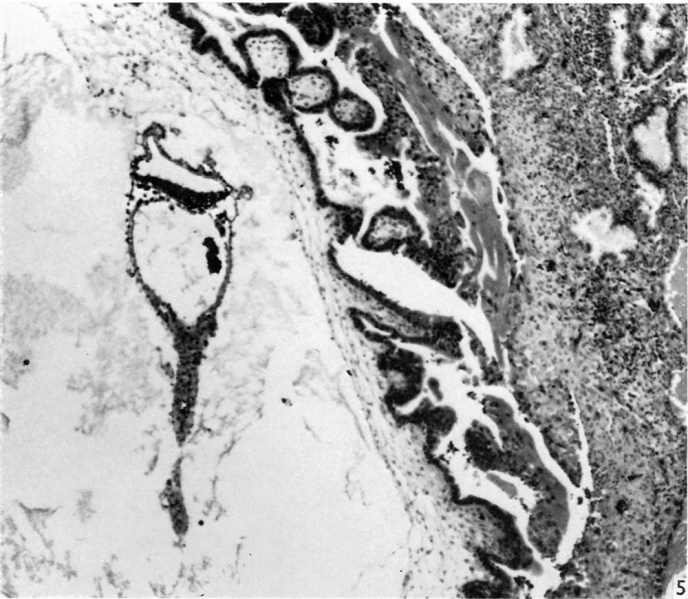
Fig. 5. Appearance of the embryo with its yolk-sac duct, and the implantation site
The yolk-sac contains a collection of erythrocytes in its lumen. The distal portion of the yolksac duct is being constricted off from the remainder. Chorionic villi in the upper part of the figure can be seen to be branching. Streamers of syncytiotrophoblast are in process of forming a trophoblastic shell. Some degeneration of the uterine glands is visible. x 71.
Reference
Lewis BV. and Harrison RG. A presomite human embryo showing a yolk-sac duct. (1966) J Anat. 100(2): 389-96. PMID: 5954785
File history
Click on a date/time to view the file as it appeared at that time.
| Date/Time | Thumbnail | Dimensions | User | Comment | |
|---|---|---|---|---|---|
| current | 23:18, 6 August 2017 | | 1,000 × 871 (184 KB) | Z8600021 (talk | contribs) | Fig. 5. Appearance of the embryo with its yolk-sac duct, and the implantation site. The yolk-sac contains a collection of erythrocytes in its lumen. The distal portion of the yolksac duct is being constricted off from the remainder. Chorionic villi in... |
You cannot overwrite this file.
File usage
The following page uses this file: