File:LewisHarrison1966 fig04.jpg
From Embryology
Size of this preview: 307 × 598 pixels.
Original file (800 × 1,559 pixels, file size: 224 KB, MIME type: image/jpeg)
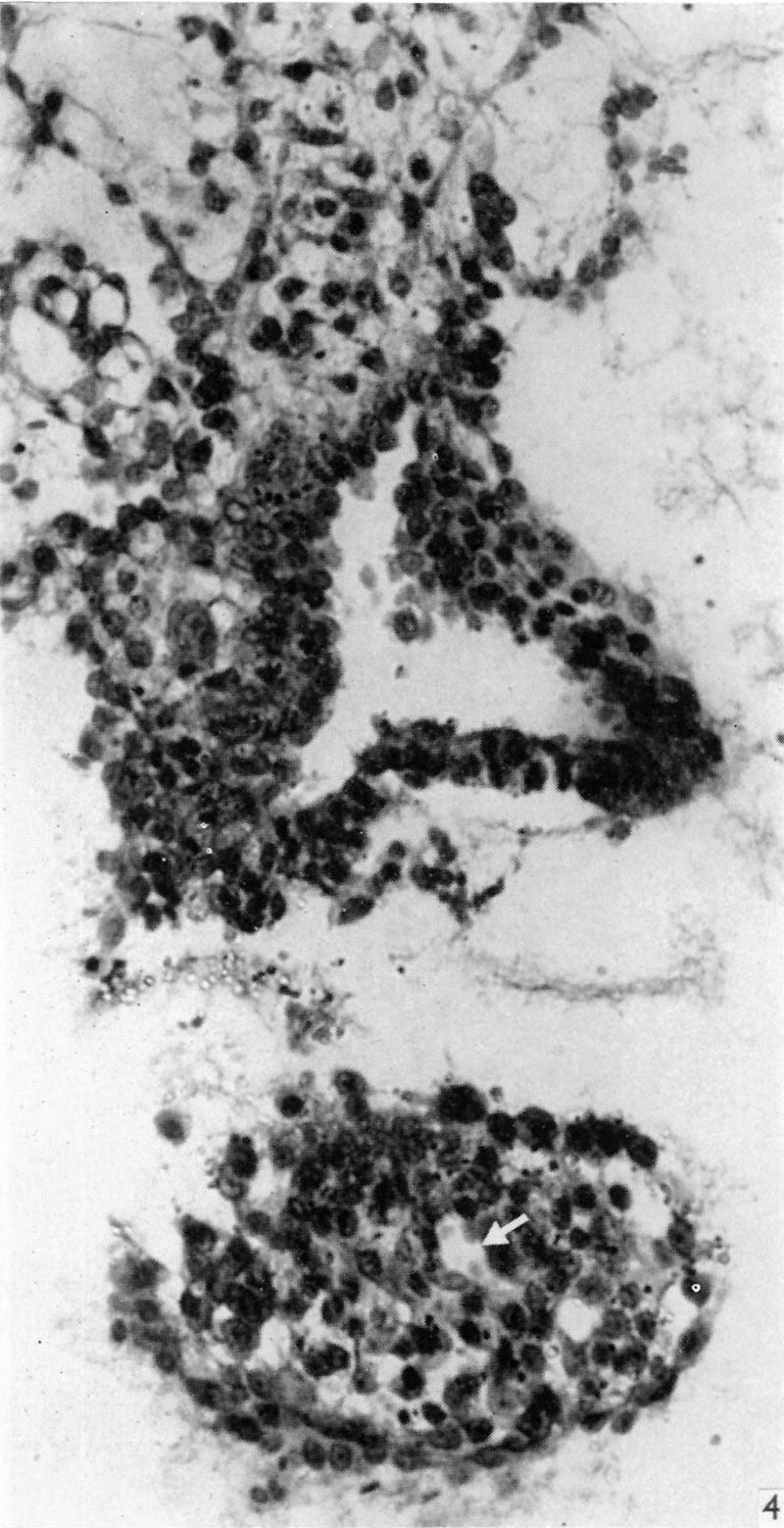
Fig. 4. Section through the embryo caudal to that in Fig. 3. The amniotic cavity is separated from the portion of the connecting stalk attached to the yolk-sac, seen in the lower part of the figure, which contains the allantoic diverticulum, indicated by an arrow. x320.
Reference
Lewis BV. and Harrison RG. A presomite human embryo showing a yolk-sac duct. (1966) J Anat. 100(2): 389-96. PMID: 5954785
File history
Click on a date/time to view the file as it appeared at that time.
| Date/Time | Thumbnail | Dimensions | User | Comment | |
|---|---|---|---|---|---|
| current | 23:14, 6 August 2017 | | 800 × 1,559 (224 KB) | Z8600021 (talk | contribs) | Fig. 4. Section through the embryo caudal to that in Fig. 3. The amniotic cavity is separated from the portion of the connecting stalk attached to the yolk-sac, seen in the lower part of the figure, which contains the allantoic diverticulum, indicated... |
You cannot overwrite this file.
File usage
The following page uses this file: