File:Lewis1909 fig06.jpg
Original file (1,000 × 991 pixels, file size: 110 KB, MIME type: image/jpeg)
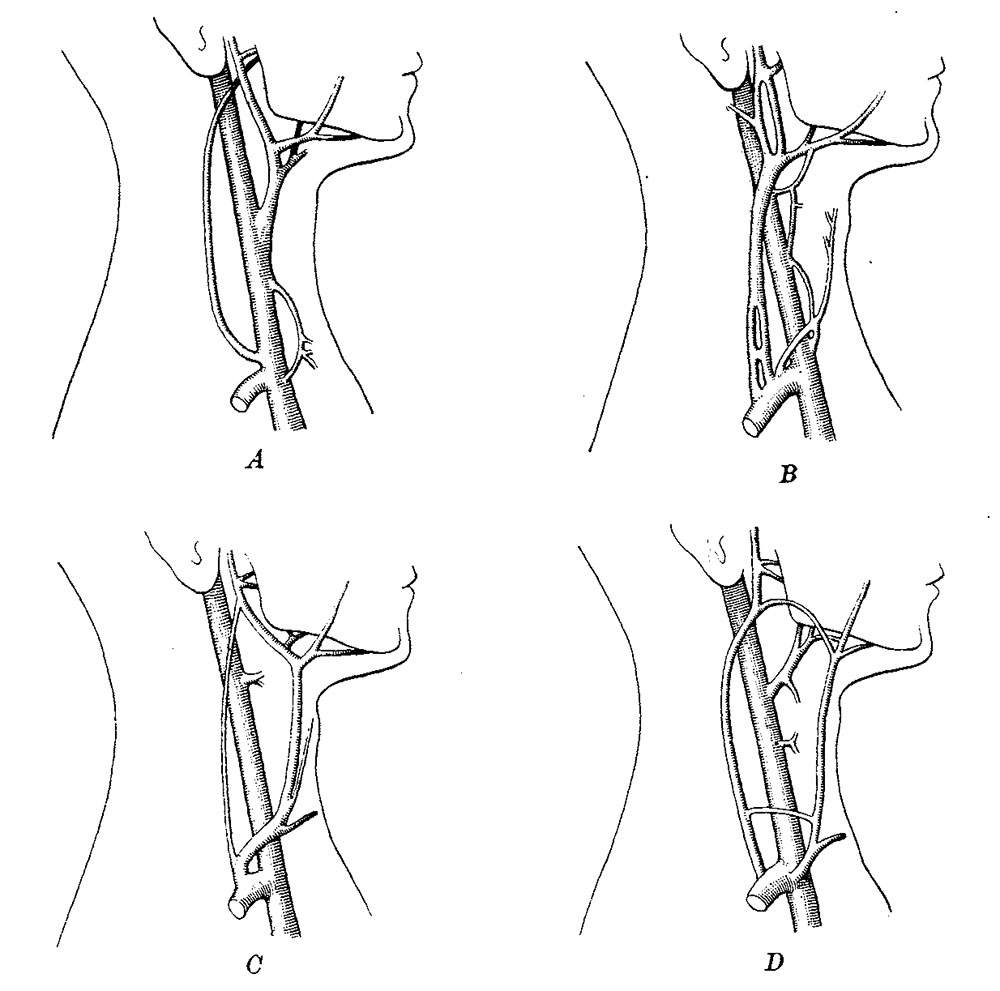
Fig. 6. The relation of the linguo-facial vein to the jugular Veins in the adult
(From dissections.)
A. The primary relation. The linguo—facial vein is a branch of the internal jugular; its submental, anterior facial, lingual, and posterior facial branches are shown in the drawing. The 1inguo—facial vein has only small anastomoses with the external jugular vein, and none with the anterior jugular, the latter vessel being scarcely represented in this case. The similarity to the embryonic relations shown in Fig. 4 is apparent.
B. The linguo—facial vein has been tapped by the external jugular so that its branches appear to belong to the latter.
C. The linguo-facial vein is drained chiefly by the anterior jugular, to which its branches appear to belong.
D. The linguo~facial vein is subdivided, so that its posterior facial branch empties into the external jugular, its anterior facial branch empties into the anterior jugular, and the lingual branch remains as a tributary of the internal jugular anomaly to be described. At the outlet of the subclavian vein there are several branches, and among them the anterior jugular vein can be identified.
| Historic Disclaimer - information about historic embryology pages |
|---|
 |
- Links: fig 1 | fig 2 | fig 3 | fig 4 | fig 5 | fig 6 | 1909 Lewis | Harvard Collection | 1905 HEC | Historic Embryology Papers | Cardiovascular System Development
Reference
Lewis FT. On the cervical veins and lymphatics in four human embryos, with an interpretation of anomalies on the subclavian and jugular veins in the adult. (1909)
Cite this page: Hill, M.A. (2024, April 26) Embryology Lewis1909 fig06.jpg. Retrieved from https://embryology.med.unsw.edu.au/embryology/index.php/File:Lewis1909_fig06.jpg
- © Dr Mark Hill 2024, UNSW Embryology ISBN: 978 0 7334 2609 4 - UNSW CRICOS Provider Code No. 00098G
File history
Click on a date/time to view the file as it appeared at that time.
| Date/Time | Thumbnail | Dimensions | User | Comment | |
|---|---|---|---|---|---|
| current | 13:44, 19 January 2017 | | 1,000 × 991 (110 KB) | Z8600021 (talk | contribs) | |
| 13:43, 19 January 2017 |  | 1,370 × 2,033 (421 KB) | Z8600021 (talk | contribs) | ==Fig. 6. == {{Lewis1909 figures}} |
You cannot overwrite this file.