File:Lewis1903 plate02L.jpg

Original file (1,846 × 2,665 pixels, file size: 287 KB, MIME type: image/jpeg)
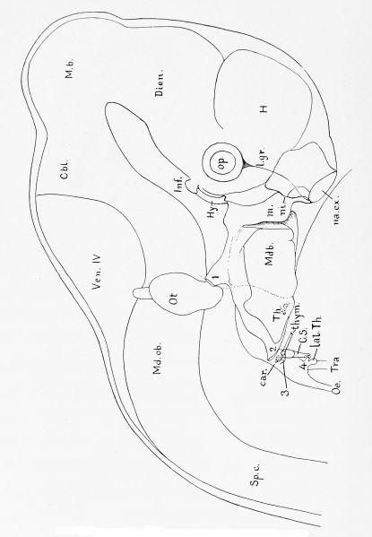
Plate II
Pig Embryo of 12.0 mm. Reconstruction from transverse sections, Series 5.
The embryo has been drawn as if transparent to show the form of its pharj'^nx, and the relations of the pharyngeal gill pouches to the grooves on the outer surface of the embryo, car. Carotid gland. Cbl, Cerebellum. CS, Cervical sinus. Dieii, Diencephalon. H, Cerebral hemisphere. Hi/, Hypophysis, liif. Infundibular gland. Lat. Th, Lateral thj^roid. 1. gr. Lachrymal groove, m. Maxillary process. M. b. Mid-brain. Mdb, Mandibular process. Md. ob. Medulla oblongata, na. ex. External naris. ni, Internal naris, closed by an epithelial plate. Oe, Oesophagvis. Op, Eye. Ot, Otocyst. Sp. c. Spinal cord. Th, Median thyroid gland, thym. Thymus. Tra, Trachea. 1, 2, 3, Ji, Entodermal pouches of the corresponding gill clefts. x 20 diams.
Reference
Lewis FT. The gross anatomy of a 12 mm pig. (1903) Amer. J Anat. 2: 221-225.
Cite this page: Hill, M.A. (2024, April 28) Embryology Lewis1903 plate02L.jpg. Retrieved from https://embryology.med.unsw.edu.au/embryology/index.php/File:Lewis1903_plate02L.jpg
- © Dr Mark Hill 2024, UNSW Embryology ISBN: 978 0 7334 2609 4 - UNSW CRICOS Provider Code No. 00098G
File history
Click on a date/time to view the file as it appeared at that time.
| Date/Time | Thumbnail | Dimensions | User | Comment | |
|---|---|---|---|---|---|
| current | 13:39, 2 August 2019 | | 1,846 × 2,665 (287 KB) | Z8600021 (talk | contribs) | |
| 13:39, 2 August 2019 |  | 1,846 × 2,789 (313 KB) | Z8600021 (talk | contribs) | ||
| 13:38, 2 August 2019 | 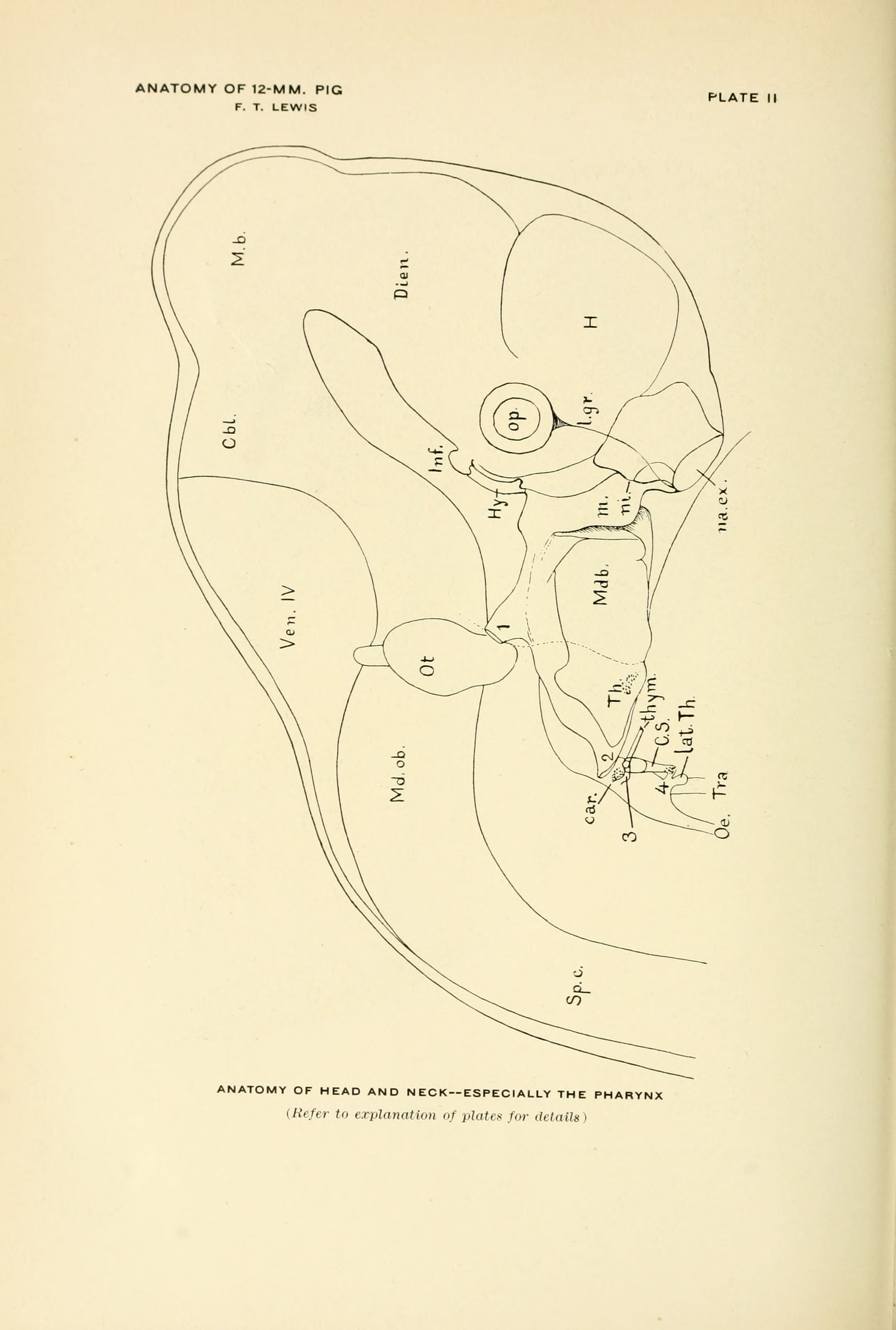 | 2,454 × 3,641 (418 KB) | Z8600021 (talk | contribs) |
You cannot overwrite this file.
File usage
The following page uses this file: