File:LeeHalpert1932 plate03.jpg

Original file (1,242 × 2,066 pixels, file size: 251 KB, MIME type: image/jpeg)
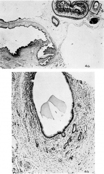
Plate 3 Fetal Gall Bladder
4 gallbladder of a 200 mm fetus in longitudinal section.
The folds of Heister and the curves of the neck assume definite forms. The viscus is the miniature of that of the newborn (a). The ductus choledochus is quite well formed. The mucosa shows numerous out-pouchings.
The mesodermal portion of the duct is composed of young connective tissue (b). Photornicrographs, (a), X 12; (b), X 120.
Reference
Halpert B. and Lee H. The gall bladder and the extrahepatic biliary passages in late embryonic and early fetal life. (1932) Anat. Rec. 54(1): 29-42.
Cite this page: Hill, M.A. (2024, April 27) Embryology LeeHalpert1932 plate03.jpg. Retrieved from https://embryology.med.unsw.edu.au/embryology/index.php/File:LeeHalpert1932_plate03.jpg
- © Dr Mark Hill 2024, UNSW Embryology ISBN: 978 0 7334 2609 4 - UNSW CRICOS Provider Code No. 00098G
File history
Click on a date/time to view the file as it appeared at that time.
| Date/Time | Thumbnail | Dimensions | User | Comment | |
|---|---|---|---|---|---|
| current | 15:38, 29 August 2017 |  | 1,242 × 2,066 (251 KB) | Z8600021 (talk | contribs) | |
| 15:26, 29 August 2017 |  | 1,366 × 2,292 (222 KB) | Z8600021 (talk | contribs) | ===Reference=== {{Ref-LeeHalpert1932}} {{Footer}} Category:Gall BladderCategory:1930's |
You cannot overwrite this file.
File usage
There are no pages that use this file.