File:Kuntz1920 fig30.jpg
From Embryology
Size of this preview: 450 × 600 pixels. Other resolution: 637 × 849 pixels.
Original file (637 × 849 pixels, file size: 110 KB, MIME type: image/jpeg)
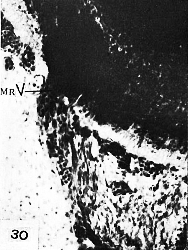
Fig. 30 Human embryo 14.5 mm in length
1267—8—2—3 x 3l5. Section through motor root of trigcminal nerve (MRV) showing migrant medullary cells.
Reference
Kuntz A. The development of the sympathetic nervous system in man. (1920) J. Comp. Neurol. 32(2): 173-229.
Cite this page: Hill, M.A. (2024, April 27) Embryology Kuntz1920 fig30.jpg. Retrieved from https://embryology.med.unsw.edu.au/embryology/index.php/File:Kuntz1920_fig30.jpg
- © Dr Mark Hill 2024, UNSW Embryology ISBN: 978 0 7334 2609 4 - UNSW CRICOS Provider Code No. 00098G
File history
Click on a date/time to view the file as it appeared at that time.
| Date/Time | Thumbnail | Dimensions | User | Comment | |
|---|---|---|---|---|---|
| current | 20:37, 28 March 2017 | | 637 × 849 (110 KB) | Z8600021 (talk | contribs) |
You cannot overwrite this file.
File usage
The following 2 pages use this file: