File:Kramer1942 fig09.jpg
Original file (1,000 × 832 pixels, file size: 120 KB, MIME type: image/jpeg)
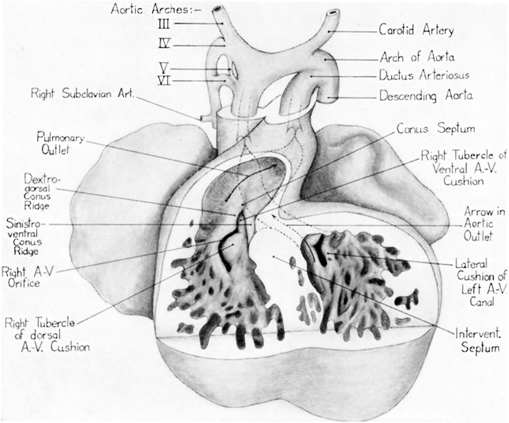
Fig. 9 Ventral view of a reconstruction of the heart of a 13 mm embryo
(Carnegie Emb. Coll. no. 841, X 42.5). Ventral wall has been removed to show the relations of the right tubercles of the dorsal and ventral atrioventricular canal cushions with the conus ridges.
| Historic Disclaimer - information about historic embryology pages |
|---|
 |
- Links:
Reference
Kramer TC. The partitioning of the truncus and conus and the formation of the membranous portion of the interventricular septum in the human heart. (1942) Amer. J Anat. 71(3): 343-370.
Cite this page: Hill, M.A. (2024, April 28) Embryology Kramer1942 fig09.jpg. Retrieved from https://embryology.med.unsw.edu.au/embryology/index.php/File:Kramer1942_fig09.jpg
- © Dr Mark Hill 2024, UNSW Embryology ISBN: 978 0 7334 2609 4 - UNSW CRICOS Provider Code No. 00098G
File history
Click on a date/time to view the file as it appeared at that time.
| Date/Time | Thumbnail | Dimensions | User | Comment | |
|---|---|---|---|---|---|
| current | 11:31, 4 February 2017 | | 1,000 × 832 (120 KB) | Z8600021 (talk | contribs) | |
| 11:29, 4 February 2017 | 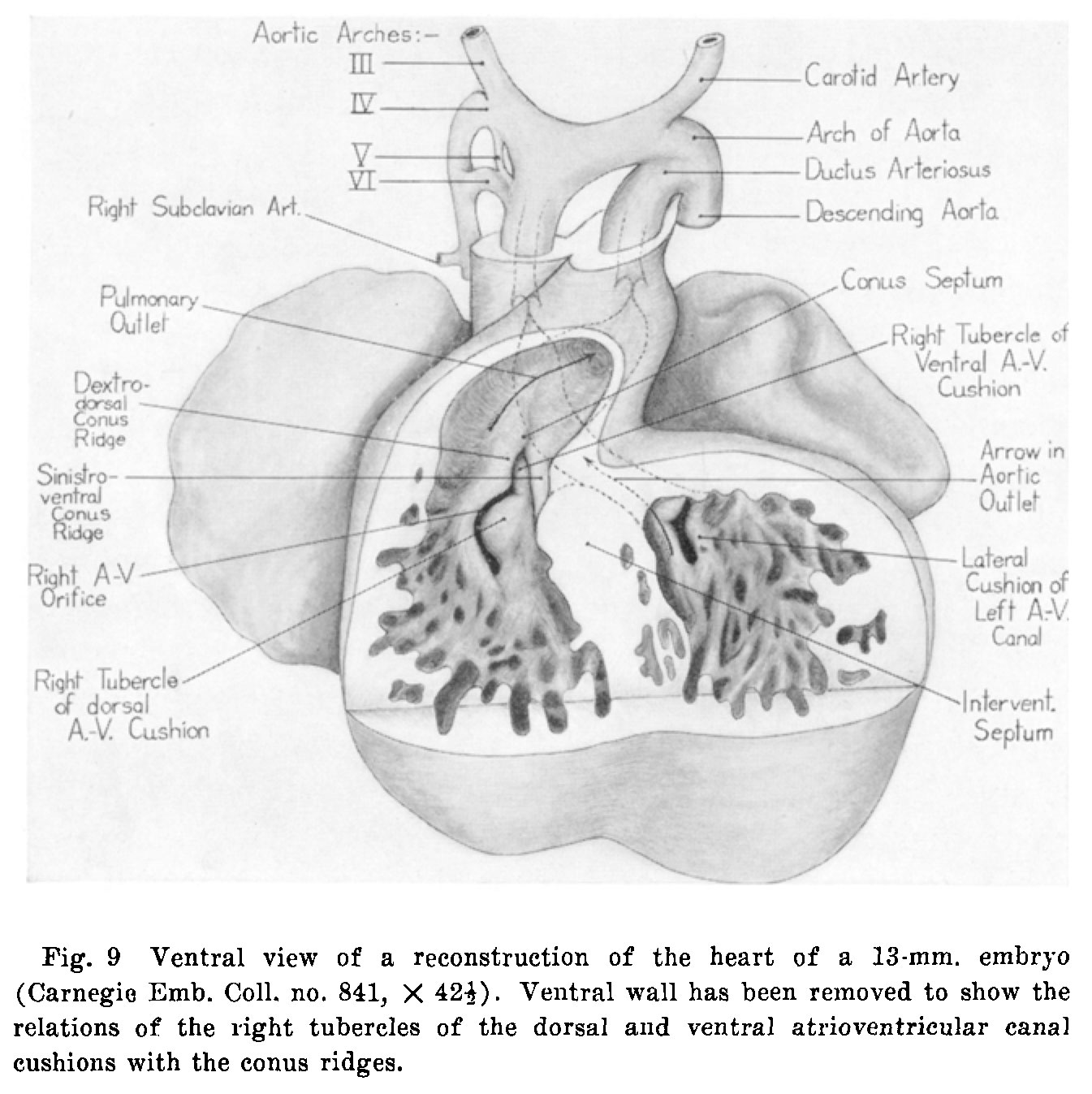 | 1,345 × 1,347 (226 KB) | Z8600021 (talk | contribs) | {{Kramer1942 figures}} |
You cannot overwrite this file.
File usage
The following page uses this file: