File:Kollmann723.jpg
Original file (737 × 611 pixels, file size: 81 KB, MIME type: image/jpeg)
- This text is a Google translate computer generated translation and may contain many errors.
Images from - Atlas of the Development of Man (Volume 2)
(Handatlas der entwicklungsgeschichte des menschen)
- Kollmann Atlas 2: Gastrointestinal | Respiratory | Urogenital | Cardiovascular | Neural | Integumentary | Smell | Vision | Hearing | Kollmann Atlas 1 | Kollmann Atlas 2 | Julius Kollmann
- Links: Julius Kollman | Atlas Vol.1 | Atlas Vol.2 | Embryology History
| Historic Disclaimer - information about historic embryology pages |
|---|
 |
Reference
Kollmann JKE. Atlas of the Development of Man (Handatlas der entwicklungsgeschichte des menschen). (1907) Vol.1 and Vol. 2. Jena, Gustav Fischer. (1898).
Cite this page: Hill, M.A. (2024, April 28) Embryology Kollmann723.jpg. Retrieved from https://embryology.med.unsw.edu.au/embryology/index.php/File:Kollmann723.jpg
- © Dr Mark Hill 2024, UNSW Embryology ISBN: 978 0 7334 2609 4 - UNSW CRICOS Provider Code No. 00098G
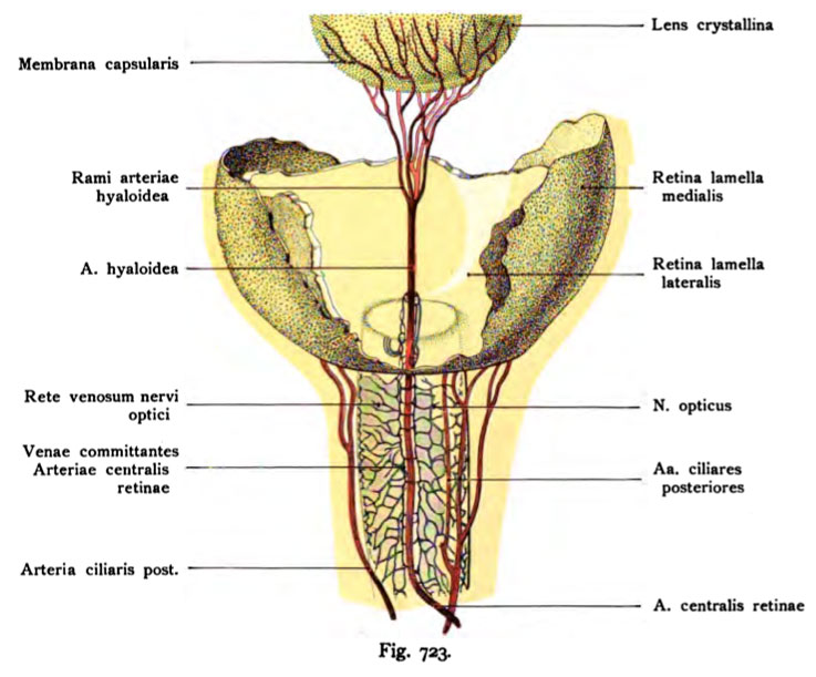
Fig. 723. Qefäfie des Nervus opticus, der Verlauf der Arteria ceotralis retinae
und der Arteria hyaloidea bei einem menschlichen Fetus von lO cm Länge.
(Nach Versari.)
Die Fortsetzung der Arteria centralis retinae durch den Glaskörper nach der Linse heißt A. hyaloidea. Eine mit der Arteria centralis retinae verlaufende Vena centralis retinae fehlt noch. Statt dessen finden sich zwei kleine Venen, Venae committantes , die sich später erst zu einer Vena centralis retinae ver- einigen. Die Venae committantes verlaufen in dem Bindegewebe, das die Arteria hyaloidea umgibt und stehen in Verbindung mit einem feinen Gefäß- netz, das sich zwischen den Bündeln des Nervus opticus befindet. Bei mensch- lichen Embryonen dieser Größe ist die Retina noch ohne Gefäße (anangisch). Die Arteria centralis retinae geht noch durch die Papilla nervi optici hindurch- ohne Zweige an die Retina abzugeben.
File history
Click on a date/time to view the file as it appeared at that time.
| Date/Time | Thumbnail | Dimensions | User | Comment | |
|---|---|---|---|---|---|
| current | 10:52, 21 October 2011 | | 737 × 611 (81 KB) | S8600021 (talk | contribs) | {{Kollmann1907}} Category:Vision Fig. 723. Qefäfie des Nervus opticus, der Verlauf der Arteria ceotralis retinae und der Arteria hyaloidea bei einem menschlichen Fetus von lO cm Länge. (Nach Versari.) Die Fortsetzung der Arteria centralis |
You cannot overwrite this file.
File usage
The following page uses this file: