File:Kollmann714.jpg
Kollmann714.jpg (625 × 553 pixels, file size: 54 KB, MIME type: image/jpeg)
- This text is a Google translate computer generated translation and may contain many errors.
Images from - Atlas of the Development of Man (Volume 2)
(Handatlas der entwicklungsgeschichte des menschen)
- Kollmann Atlas 2: Gastrointestinal | Respiratory | Urogenital | Cardiovascular | Neural | Integumentary | Smell | Vision | Hearing | Kollmann Atlas 1 | Kollmann Atlas 2 | Julius Kollmann
- Links: Julius Kollman | Atlas Vol.1 | Atlas Vol.2 | Embryology History
| Historic Disclaimer - information about historic embryology pages |
|---|
 |
Reference
Kollmann JKE. Atlas of the Development of Man (Handatlas der entwicklungsgeschichte des menschen). (1907) Vol.1 and Vol. 2. Jena, Gustav Fischer. (1898).
Cite this page: Hill, M.A. (2024, April 28) Embryology Kollmann714.jpg. Retrieved from https://embryology.med.unsw.edu.au/embryology/index.php/File:Kollmann714.jpg
- © Dr Mark Hill 2024, UNSW Embryology ISBN: 978 0 7334 2609 4 - UNSW CRICOS Provider Code No. 00098G
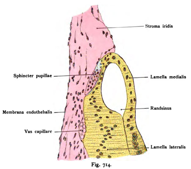
Fig. 714. Entwicklung des Sphincter pupillae.
Frühe Stufe. Radiärschnitt durch die Iris eines 10,2 cm langen menschlichen Fetus (Gesamtlänge.) (5. Monat.) Nach Beseitigung des Pigmentes.
(Nach Szili jun.)
Vergl. die Fig. 713, den Vertikalschnitt der vorderen Augenhälfte eines menschlichen Embryo von 8—9 Wochen behufs leichterer Orientierung. Der bindegewebige Anteil der Iris, Stroma iridis, bedeckt den epithelialen Anteil. Die laterale und mediale Lamelle des Augenbechers sind künstlich befreit von Pigment und in der nämlichen Farbe dargestellt. Am lateralen Rande gehen die beiden Lamellen ineinander über und umschließen einen ovalen Ringsinus, der später verschwindet. An der Übergangsstelle der beiden Lamellen erhebt sich ein kurzer, flächenhaft über die laterale Lamelle gelagerter Fortsatz, die Anlage des Sphincter pupillae aus den Zellen der Umschlagstelle. Der Fortsatz ist gegen die Lamella lateralis durch eine helle Furche abgegrenzt
File history
Click on a date/time to view the file as it appeared at that time.
| Date/Time | Thumbnail | Dimensions | User | Comment | |
|---|---|---|---|---|---|
| current | 10:48, 21 October 2011 | | 625 × 553 (54 KB) | S8600021 (talk | contribs) | {{Kollmann1907}} Category:Vision Fig. 714. Entwicklung des Sphincter pupillae. Frühe Stufe. Radiärschnitt durch die Iris eines 10,2 cm langen menschlichen Fetus (Gesamtlänge.) (5. Monat.) Nach Beseitigung des Pigmentes. (Nach Szili jun.) |
You cannot overwrite this file.
File usage
The following page uses this file: