File:Kollmann696.jpg
Kollmann696.jpg (685 × 494 pixels, file size: 40 KB, MIME type: image/jpeg)
- This text is a Google translate computer generated translation and may contain many errors.
Images from - Atlas of the Development of Man (Volume 2)
(Handatlas der entwicklungsgeschichte des menschen)
- Kollmann Atlas 2: Gastrointestinal | Respiratory | Urogenital | Cardiovascular | Neural | Integumentary | Smell | Vision | Hearing | Kollmann Atlas 1 | Kollmann Atlas 2 | Julius Kollmann
- Links: Julius Kollman | Atlas Vol.1 | Atlas Vol.2 | Embryology History
| Historic Disclaimer - information about historic embryology pages |
|---|
 |
Reference
Kollmann JKE. Atlas of the Development of Man (Handatlas der entwicklungsgeschichte des menschen). (1907) Vol.1 and Vol. 2. Jena, Gustav Fischer. (1898).
Cite this page: Hill, M.A. (2024, April 27) Embryology Kollmann696.jpg. Retrieved from https://embryology.med.unsw.edu.au/embryology/index.php/File:Kollmann696.jpg
- © Dr Mark Hill 2024, UNSW Embryology ISBN: 978 0 7334 2609 4 - UNSW CRICOS Provider Code No. 00098G
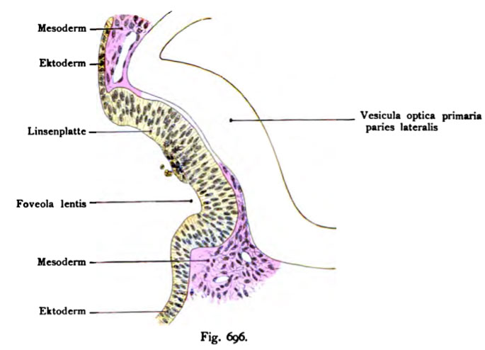
Fig. 696. Linseoanlage bei einem lOtäsigen Kaninchenembryo.
(Nach Rabl.)
Die laterale Wand der primären Augenblase ist konkav. In der Ver- tiefung liegt jetzt das Linsengrübchen, Foveola lentis. Auf dem Boden des Grüb- chens liegt ein Zellhaufen, unter ihm dicht nebeneinander Teilungsfiguren. Lateral vom Zellhaufen einige kugelige Gebilde. Die Linsenplatte erweist sich auch noch auf dieser Entwicklungsstufe als eine direkte Fortsetzung des Ektoderms. Das Mesoderm dringt zwischen Linsenplatte und der lateralen Wand der Augen- blase mehr und mehr gegen die Mitte des Linsengrübchens (Fossula lentis) vor.
File history
Click on a date/time to view the file as it appeared at that time.
| Date/Time | Thumbnail | Dimensions | User | Comment | |
|---|---|---|---|---|---|
| current | 10:39, 21 October 2011 | | 685 × 494 (40 KB) | S8600021 (talk | contribs) | {{Kollmann1907}} Category:Vision Fig. 696. Linseoanlage bei einem lOtäsigen Kaninchenembryo. (Nach Rabl.) Die laterale Wand der primären Augenblase ist konkav. In der Ver- tiefung liegt jetzt das Linsengrübchen, Foveola lentis. Auf dem Bo |
You cannot overwrite this file.
File usage
The following page uses this file: