File:Kollmann680.jpg

Original file (738 × 660 pixels, file size: 66 KB, MIME type: image/jpeg)
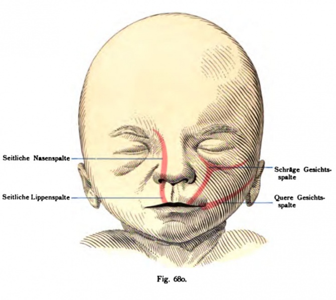
Fig. 680. Face with the system of embryonic splitting
The fork-shaped top column, the lower leg of the oral cavity starts, while the two upper leg soft at the lower eyelid apart, can remain open, and then provides the so-called "oblique face - column" dar. (compare Fig. 677) In very severe cases, it surrounds the eye from below. - The "transverse facial cleft" corresponds to an open-remain the gaps between the maxillary process and the Mandibular the First branchial arch. (Compare Fig. 674) - By far the most common Hemmimgs training is the (harelip). It goes without exception go-to nostril. (Compare Fig. 675), it extends to even higher up to the olfactory region, it is called "divisive lateral nose." (See the Fig. 675), a media column in the upper lip is formed by the permanent separation of the process of globular which are otherwise due to the formation of adhesions so-called middle lip (labium medianum) and takes part of the philtrum. (cf. Fig. 677) In severe cases, even the lack of association-tion of the nose. A division of the lower lip rests on the permanent separation of the mandibular arch (rarely). (cf. Fig. 345)
- This text is a Google translate computer generated translation and may contain many errors.
Images from - Atlas of the Development of Man (Volume 2)
(Handatlas der entwicklungsgeschichte des menschen)
- Kollmann Atlas 2: Gastrointestinal | Respiratory | Urogenital | Cardiovascular | Neural | Integumentary | Smell | Vision | Hearing | Kollmann Atlas 1 | Kollmann Atlas 2 | Julius Kollmann
- Links: Julius Kollman | Atlas Vol.1 | Atlas Vol.2 | Embryology History
| Historic Disclaimer - information about historic embryology pages |
|---|
| Pages where the terms "Historic" (textbooks, papers, people, recommendations) appear on this site, and sections within pages where this disclaimer appears, indicate that the content and scientific understanding are specific to the time of publication. This means that while some scientific descriptions are still accurate, the terminology and interpretation of the developmental mechanisms reflect the understanding at the time of original publication and those of the preceding periods, these terms, interpretations and recommendations may not reflect our current scientific understanding. (More? Embryology History | Historic Embryology Papers) |
Reference
Kollmann JKE. Atlas of the Development of Man (Handatlas der entwicklungsgeschichte des menschen). (1907) Vol.1 and Vol. 2. Jena, Gustav Fischer. (1898).
Cite this page: Hill, M.A. (2024, April 28) Embryology Kollmann680.jpg. Retrieved from https://embryology.med.unsw.edu.au/embryology/index.php/File:Kollmann680.jpg
- © Dr Mark Hill 2024, UNSW Embryology ISBN: 978 0 7334 2609 4 - UNSW CRICOS Provider Code No. 00098G
Fig. 680. Gesicht mit dem einsezeichoeten System der Embryonalspalten.
Die nach oben gabelförmige Spalte, deren unterer Schenkel von der Mund-
höhle ausgeht, während die beiden oberen Schenkel am unteren Lid auseinander
weichen, kann offen bleiben und stellt dann die sog. „schräge Gesichts -
spalte" dar. (Vergl. die Fig. 677.) In ganz schweren Fällen umgreift sie das
Auge von unten her. — Die „quere Gesichtspalte" entspricht einem Offen-
bleiben der Spalte zwischen dem Oberkieferfortsatz und dem Mandibularteil des
I. Kiemenbogens. (Vergl. die Fig. 674.) — Weitaus die häufigste Hemmimgs-
bildung ist die „s e i 1 1 i c h e Li p p e n s p a 1 1 e (Hasenscharte). Sie geht ausnahms-
los bis zum Nasenloch. (Vergl. die Fig. 675.) Erstreckt sie sich noch höher
hinauf bis zur Regio olfactoria, so heißt sie „seitliche Nasen spalte". (Vergl.
die Fig. 675.) Eine Medianspalte der Oberlippe entsteht durch die
bleibende Trennung der Processus globulares, welche sonst durch Verwachsung
an der Bildung der sog. Mittellippe (Labium medianum) und des Philtrum be-
teiligt sind. (Vergl. die Fig. 677.) In schweren Fällen fehlt selbst die Vereini-
gung zur Nasenspitze. Eine Spaltung der Unterlippe beruht auf der bleibenden
Trennung des Mandibularbogens (selten). (Vergl. Fig. 345.)
File history
Click on a date/time to view the file as it appeared at that time.
| Date/Time | Thumbnail | Dimensions | User | Comment | |
|---|---|---|---|---|---|
| current | 10:08, 21 October 2011 |  | 738 × 660 (66 KB) | S8600021 (talk | contribs) | {{Kollmann1907}} Category:Smell Fig. 680. Gesicht mit dem einsezeichoeten System der Embryonalspalten. Die nach oben gabelförmige Spalte, deren unterer Schenkel von der Mund- höhle ausgeht, während die beiden oberen Schenkel am unteren Lid a |
You cannot overwrite this file.
File usage
The following page uses this file:
