File:Kollmann679.jpg
Kollmann679.jpg (727 × 450 pixels, file size: 55 KB, MIME type: image/jpeg)
- This text is a Google translate computer generated translation and may contain many errors.
Images from - Atlas of the Development of Man (Volume 2)
(Handatlas der entwicklungsgeschichte des menschen)
- Kollmann Atlas 2: Gastrointestinal | Respiratory | Urogenital | Cardiovascular | Neural | Integumentary | Smell | Vision | Hearing | Kollmann Atlas 1 | Kollmann Atlas 2 | Julius Kollmann
- Links: Julius Kollman | Atlas Vol.1 | Atlas Vol.2 | Embryology History
| Historic Disclaimer - information about historic embryology pages |
|---|
 |
Reference
Kollmann JKE. Atlas of the Development of Man (Handatlas der entwicklungsgeschichte des menschen). (1907) Vol.1 and Vol. 2. Jena, Gustav Fischer. (1898).
Cite this page: Hill, M.A. (2024, April 28) Embryology Kollmann679.jpg. Retrieved from https://embryology.med.unsw.edu.au/embryology/index.php/File:Kollmann679.jpg
- © Dr Mark Hill 2024, UNSW Embryology ISBN: 978 0 7334 2609 4 - UNSW CRICOS Provider Code No. 00098G
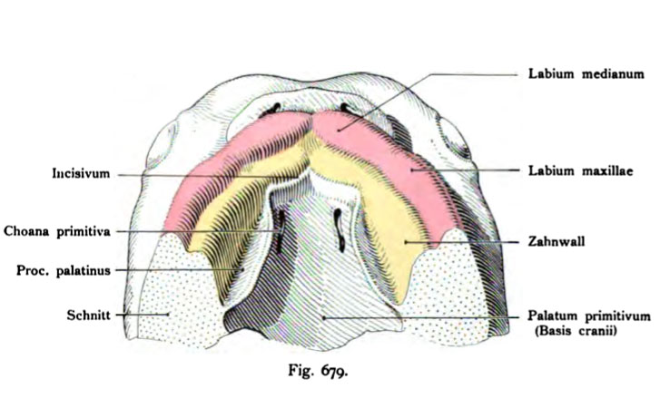
Fig. 679. Ventrale Umgebung des primitiven Gaumens
bei einem menschlichen Embryo von etwa 19 mm Länge. (Etwa 15 mal vergr.)
(Anatomische Sammlung in Basel)
Die Lippen beginnen sich durch eine epitheliale Einsenkung von der Anlage des Zwischenkiefers und von dem übrigen Kieferrand zu trennen. Das Mittel- stück der Lippen und das Incisivum entstehen durch Vereinigung der beiden Processus globulares (vergl. Fig. 675). Der Oberkieferfortsatz liefert die „Ober- kieferlippe", sein mittlerer Abschnitt den Zahnwall, der sich auch vom Incisivum abgliedert. Damit ist sowohl die Facies palatina des Incisivum, als die Facies palatina des Processus palatinus abgegrenzt. Diese letzterwähnten Platten des sekundären Gaumens treffen sich später in der Mittellinie, in der Sutura palatina. Allein jetzt, bei 25 — 30 mm Scheitelsteißlänge sind sie noch gegen den Unterkiefer, also kaudal gerichtet und in den Raum zwischen die Processus palatini ragt die Zunge tief hinein (vergl. die Figg. 274 und 276, Entwicklung des Gaumens in dem Abschnitt „Embryologia ossium".
File history
Click on a date/time to view the file as it appeared at that time.
| Date/Time | Thumbnail | Dimensions | User | Comment | |
|---|---|---|---|---|---|
| current | 10:07, 21 October 2011 | | 727 × 450 (55 KB) | S8600021 (talk | contribs) | {{Kollmann1907}} Category:Smell Fig. 679. Ventrale Umgebung des primitiven Gaumens bei einem menschlichen Embryo von etwa 19 mm Länge. (Etwa 15 mal vergr.) (Anatomische Sammlung in Basel) Die Lippen beginnen sich durch eine epitheliale Ein |
You cannot overwrite this file.
File usage
The following page uses this file: