File:Kollmann671.jpg
Kollmann671.jpg (686 × 354 pixels, file size: 40 KB, MIME type: image/jpeg)
- This text is a Google translate computer generated translation and may contain many errors.
Images from - Atlas of the Development of Man (Volume 2)
(Handatlas der entwicklungsgeschichte des menschen)
- Kollmann Atlas 2: Gastrointestinal | Respiratory | Urogenital | Cardiovascular | Neural | Integumentary | Smell | Vision | Hearing | Kollmann Atlas 1 | Kollmann Atlas 2 | Julius Kollmann
- Links: Julius Kollman | Atlas Vol.1 | Atlas Vol.2 | Embryology History
| Historic Disclaimer - information about historic embryology pages |
|---|
 |
Reference
Kollmann JKE. Atlas of the Development of Man (Handatlas der entwicklungsgeschichte des menschen). (1907) Vol.1 and Vol. 2. Jena, Gustav Fischer. (1898).
Cite this page: Hill, M.A. (2024, April 26) Embryology Kollmann671.jpg. Retrieved from https://embryology.med.unsw.edu.au/embryology/index.php/File:Kollmann671.jpg
- © Dr Mark Hill 2024, UNSW Embryology ISBN: 978 0 7334 2609 4 - UNSW CRICOS Provider Code No. 00098G
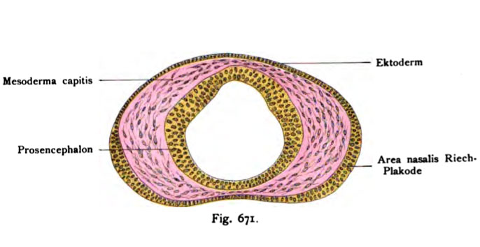
Fig. 671. Eotwickluns des Genichsorgans
bei einem menschlichen Embryo vom Anfang der 4. Woche.
(Nach Peter.)
Schnitt durch den Vorderkopf mit dem Prosencephalon und dem Kopf- mesoderm. Zu beiden Seiten findet sich wie bei allen bisher untersuchten Säugern eine Verdickung des Ektoderms: die Riechpiakode. Das Ektoderm ist im Bereich dieser Gegend mehrschichtig. Die Riechpiakode ist noch nicht scharf begrenzt, sondern setzt sich allmählich in die einfache Zellenlage der übrigen Kopfrundung fort.
File history
Click on a date/time to view the file as it appeared at that time.
| Date/Time | Thumbnail | Dimensions | User | Comment | |
|---|---|---|---|---|---|
| current | 10:01, 21 October 2011 | | 686 × 354 (40 KB) | S8600021 (talk | contribs) | {{Kollmann1907}} Category:Smell Fig. 671. Eotwickluns des Genichsorgans bei einem menschlichen Embryo vom Anfang der 4. Woche. (Nach Peter.) Schnitt durch den Vorderkopf mit dem Prosencephalon und dem Kopf- mesoderm. Zu beiden Seiten findet |
You cannot overwrite this file.
File usage
The following page uses this file: