File:Kollmann666.jpg

Original file (897 × 1,000 pixels, file size: 140 KB, MIME type: image/jpeg)
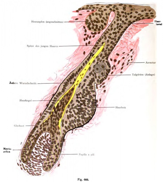
Fig. 666. Development of the wool hair (lanugo) in a human fetus of 5.5 months
(Rückenhaut.) 460 times magnified
(After St eye.)
The papilla has now grown considerably in length, consists of a hair cone whose peripheral cells to the inner root sheath, while the axial to alter the hair. The hair is yellow with the applied treatment. At this stage of development this is the young hair, because it is still in the hair sheath "infected vaginal hair. It will penetrate through extension and eventually is produced on the surface of the skin. After the failure of the wool hair (lanugo) there follows an outgoing of the cylinder bed hair cells in the formation of new cells, which extend down to the old papilla itself. The matrix produced then by the above-described mode, produces a new hair. if a hair follicle is still missing, it occurs later.
- This text is a Google translate computer generated translation and may contain many errors.
Images from - Atlas of the Development of Man (Volume 2)
(Handatlas der entwicklungsgeschichte des menschen)
- Kollmann Atlas 2: Gastrointestinal | Respiratory | Urogenital | Cardiovascular | Neural | Integumentary | Smell | Vision | Hearing | Kollmann Atlas 1 | Kollmann Atlas 2 | Julius Kollmann
- Links: Julius Kollman | Atlas Vol.1 | Atlas Vol.2 | Embryology History
| Historic Disclaimer - information about historic embryology pages |
|---|
| Pages where the terms "Historic" (textbooks, papers, people, recommendations) appear on this site, and sections within pages where this disclaimer appears, indicate that the content and scientific understanding are specific to the time of publication. This means that while some scientific descriptions are still accurate, the terminology and interpretation of the developmental mechanisms reflect the understanding at the time of original publication and those of the preceding periods, these terms, interpretations and recommendations may not reflect our current scientific understanding. (More? Embryology History | Historic Embryology Papers) |
Reference
Kollmann JKE. Atlas of the Development of Man (Handatlas der entwicklungsgeschichte des menschen). (1907) Vol.1 and Vol. 2. Jena, Gustav Fischer. (1898).
Cite this page: Hill, M.A. (2024, April 27) Embryology Kollmann666.jpg. Retrieved from https://embryology.med.unsw.edu.au/embryology/index.php/File:Kollmann666.jpg
- © Dr Mark Hill 2024, UNSW Embryology ISBN: 978 0 7334 2609 4 - UNSW CRICOS Provider Code No. 00098G
Fig. 666. Entwicklung des Wollhaares, Lanugo,
bei einem menschlichen Fetus von 5 ^/2 Monaten. (Rückenhaut.) 460 mal vergr.
(Nach St Öhr.)
Die Haarpapille ist jetzt ansehnlich in die Länge gewachsen, umfaßt vom Haarkegel, dessen periphere Zellen zur inneren Wurzelscheide werden, während die axialen zum Haar sich umändern. Das Haar ist bei der angewendeten Behandlung gelblich. Auf dieser Entwicklungsstufe heißt das junge Haar, weil es noch in der Haarscheide steckt „Scheidenhaar. Durch Verlängerung dringt es schließlich auf der Oberfläche der Haut hervor. Nach dem Ausfallen des Wollhaares folgt eine von den Zylinderzellen des Haarbettes ausgehende Neubildung von Zellen, die bis auf die alte Papille hinab sich ausdehnen. Die Matrix produziert dann nach dem eben beschriebenen Modus ein neues Haar. Ein Haarkanal fehlt noch, er tritt später auf.
File history
Click on a date/time to view the file as it appeared at that time.
| Date/Time | Thumbnail | Dimensions | User | Comment | |
|---|---|---|---|---|---|
| current | 07:38, 21 October 2011 |  | 897 × 1,000 (140 KB) | S8600021 (talk | contribs) | {{Kollmann1907}} Category:Integumentary |
You cannot overwrite this file.
File usage
The following 2 pages use this file:
