File:Kollmann586.jpg
Kollmann586.jpg (519 × 557 pixels, file size: 40 KB, MIME type: image/jpeg)
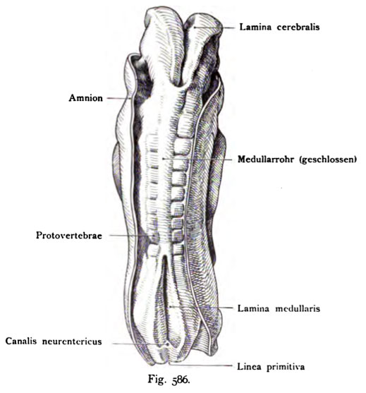
Fig. 586. The primordial central nervous system in human embryo of 2.11 mm length
Age around 13 - 14 days. Norma dorsalis. Reconstruction.
(After Eternod)
The neural tube is at the head and body still wide open and will end lamina as cerebral, cranial plate and lamina medullaris, called medullary But you can see the symmetrical halves already collected and Term to approach more and more. In the middle section of the body The neural tube is closed at the plate. At the end of the body sets the Neural plate to both sides of the neurenteric canal continues on. behind the neurenteric canal is still present the primitive groove. There are still no gills developed, not even in the embryo Fig. 585.
- This text is a Google translate computer generated translation and may contain many errors.
Images from - Atlas of the Development of Man (Volume 2)
(Handatlas der entwicklungsgeschichte des menschen)
- Kollmann Atlas 2: Gastrointestinal | Respiratory | Urogenital | Cardiovascular | Neural | Integumentary | Smell | Vision | Hearing | Kollmann Atlas 1 | Kollmann Atlas 2 | Julius Kollmann
- Links: Julius Kollman | Atlas Vol.1 | Atlas Vol.2 | Embryology History
| Historic Disclaimer - information about historic embryology pages |
|---|
| Pages where the terms "Historic" (textbooks, papers, people, recommendations) appear on this site, and sections within pages where this disclaimer appears, indicate that the content and scientific understanding are specific to the time of publication. This means that while some scientific descriptions are still accurate, the terminology and interpretation of the developmental mechanisms reflect the understanding at the time of original publication and those of the preceding periods, these terms, interpretations and recommendations may not reflect our current scientific understanding. (More? Embryology History | Historic Embryology Papers) |
Reference
Kollmann JKE. Atlas of the Development of Man (Handatlas der entwicklungsgeschichte des menschen). (1907) Vol.1 and Vol. 2. Jena, Gustav Fischer. (1898).
Cite this page: Hill, M.A. (2024, April 27) Embryology Kollmann586.jpg. Retrieved from https://embryology.med.unsw.edu.au/embryology/index.php/File:Kollmann586.jpg
- © Dr Mark Hill 2024, UNSW Embryology ISBN: 978 0 7334 2609 4 - UNSW CRICOS Provider Code No. 00098G
Fig. 586. Die Anlage des Zentralnervensystems bei eüiem menschlichen Embryo
von 2,11 mm Länge,
Alter etwa 13 — 14 Tage. Norma dorsalis. Rekonstruktion.
(Nach Eternod)
Das MeduUarrohr ist am Kopf und Körperende noch weit offen und wird als Lamina cerebralis, Hirnplatte und Lamina medullaris, MeduUarplatte be- zeichnet Man sieht jedoch die symmetrischen Hälften schon erhoben und im Begriff, sich mehr und mehr zu nähern. Im mittleren Abschnitt des Körpers ist die Platte zum MeduUarrohr geschlossen. Am Körperende setzt sich die MeduUarplatte zu beiden Seiten des CanaUs neurent6ricus weiter fort. Hinter dem Canalis neurentericus ist noch die Primitivrinne vorhanden. Es sind noch keine Kiemenbogen entwickelt, auch bei dem Embryo Fig. 585 nicht
File history
Click on a date/time to view the file as it appeared at that time.
| Date/Time | Thumbnail | Dimensions | User | Comment | |
|---|---|---|---|---|---|
| current | 16:25, 17 October 2011 |  | 519 × 557 (40 KB) | S8600021 (talk | contribs) | {{Kollmann1907}} |
You cannot overwrite this file.
File usage
The following page uses this file:

