File:Kollmann513.jpg
Kollmann513.jpg (662 × 588 pixels, file size: 58 KB, MIME type: image/jpeg)
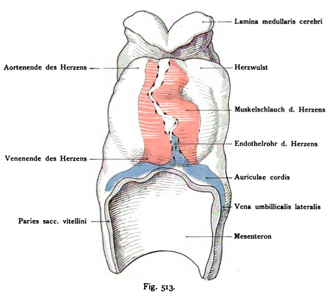
Fig. 513. Human embryo heart and heart bead of 2.11 mm in length
Age 13-14 days.
(After Eternod.) Reconstruction.
It is the front half of the embryo with the strongly projecting transparent Herzwulst shown. Inside the heart (red) of the endothelium- crossed pipe. The endothelial moves first from left to right and then rises up in the air. The heart goes up into the range of front edge close to the brain Stomadaeum (the bay mouth). the primitive Auriculae cordis are still inferior and are an extension of the oval Lateral umbilical vein dar. on the marginal ridge of the yolk sac, this Veins are also in the rabbit, there they are called veins omphalomesenterica in van Beneden and Julin Ravn. The space between the Muscle of the heart tube and the membrane of the heart is filled with lymph.
- This text is a Google translate computer generated translation and may contain many errors.
Images from - Atlas of the Development of Man (Volume 2)
(Handatlas der entwicklungsgeschichte des menschen)
- Kollmann Atlas 2: Gastrointestinal | Respiratory | Urogenital | Cardiovascular | Neural | Integumentary | Smell | Vision | Hearing | Kollmann Atlas 1 | Kollmann Atlas 2 | Julius Kollmann
- Links: Julius Kollman | Atlas Vol.1 | Atlas Vol.2 | Embryology History
| Historic Disclaimer - information about historic embryology pages |
|---|
| Pages where the terms "Historic" (textbooks, papers, people, recommendations) appear on this site, and sections within pages where this disclaimer appears, indicate that the content and scientific understanding are specific to the time of publication. This means that while some scientific descriptions are still accurate, the terminology and interpretation of the developmental mechanisms reflect the understanding at the time of original publication and those of the preceding periods, these terms, interpretations and recommendations may not reflect our current scientific understanding. (More? Embryology History | Historic Embryology Papers) |
Reference
Kollmann JKE. Atlas of the Development of Man (Handatlas der entwicklungsgeschichte des menschen). (1907) Vol.1 and Vol. 2. Jena, Gustav Fischer. (1898).
Cite this page: Hill, M.A. (2024, April 28) Embryology Kollmann513.jpg. Retrieved from https://embryology.med.unsw.edu.au/embryology/index.php/File:Kollmann513.jpg
- © Dr Mark Hill 2024, UNSW Embryology ISBN: 978 0 7334 2609 4 - UNSW CRICOS Provider Code No. 00098G
Fig. 513. Herz und Herzwulst Menschlicher Embryo von 2,11 mm Länge.
Alter 13—14 Tage.
(Nach Eternod.) Rekonstruktion.
Es ist die vordere Hälfte des Embryo mit dem stark vorspringenden durchsichtigen Herzwulst dargestellt. Im Innern das Herz (rot) von dem Endothel- rohr durchzogen. Das Endothelrohr zieht zuerst von links nach rechts und steigt dann gerade in die Höhe. Das Herz reicht hinauf in den Bereich des vorderen Hirnrandes dicht an das Stomadaeum (die Mundbucht). Die primitiven Auriculae cordis liegen noch kaudal und stellen eine ovale Erweiterung der Venae umbilicales laterales auf dem Randwulst des Dottersackes dar. Diese Venen liegen ebenso beim Kaninchen, dort heißen sie Venae omphalo-mesentericae bei van Beneden, Julin und Ravn. Der Raum zwischen dem Muskelschlauch des Herzens und der Membran des Herzwulstes ist angefüllt mit Urlymphe.
File history
Click on a date/time to view the file as it appeared at that time.
| Date/Time | Thumbnail | Dimensions | User | Comment | |
|---|---|---|---|---|---|
| current | 23:17, 16 October 2011 |  | 662 × 588 (58 KB) | S8600021 (talk | contribs) | {{Kollmann1907}} |
You cannot overwrite this file.
File usage
The following page uses this file:

