File:Kollmann445.jpg

Original file (776 × 738 pixels, file size: 112 KB, MIME type: image/jpeg)
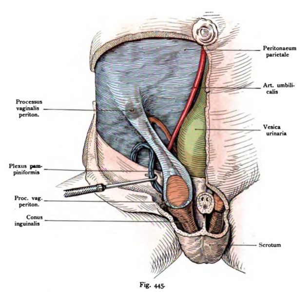
Fig. 445. The process of vaginalis peritoneai the testis and epididymis in the interior
After a fresh preparation. (Anatomical Collection in Basel)
Skin and muscles, penis, and part of the scrotum are removed and in the area of the abdomen leaving only the peritoneum. This is the Departure of the processus vaginalis peritonaei visible. The Plexus pampini- formis is pulled aside by a hook and is not of the processus enclosed vaginalis.
Moves to the median plane down behind the peritoneum the vas deferens descends into the pelvis and the bladder and the umbilical artery up to the navel.
- This text is a Google translate computer generated translation and may contain many errors.
Images from - Atlas of the Development of Man (Volume 2)
(Handatlas der entwicklungsgeschichte des menschen)
- Kollmann Atlas 2: Gastrointestinal | Respiratory | Urogenital | Cardiovascular | Neural | Integumentary | Smell | Vision | Hearing | Kollmann Atlas 1 | Kollmann Atlas 2 | Julius Kollmann
- Links: Julius Kollman | Atlas Vol.1 | Atlas Vol.2 | Embryology History
| Historic Disclaimer - information about historic embryology pages |
|---|
| Pages where the terms "Historic" (textbooks, papers, people, recommendations) appear on this site, and sections within pages where this disclaimer appears, indicate that the content and scientific understanding are specific to the time of publication. This means that while some scientific descriptions are still accurate, the terminology and interpretation of the developmental mechanisms reflect the understanding at the time of original publication and those of the preceding periods, these terms, interpretations and recommendations may not reflect our current scientific understanding. (More? Embryology History | Historic Embryology Papers) |
Reference
Kollmann JKE. Atlas of the Development of Man (Handatlas der entwicklungsgeschichte des menschen). (1907) Vol.1 and Vol. 2. Jena, Gustav Fischer. (1898).
Cite this page: Hill, M.A. (2024, April 28) Embryology Kollmann445.jpg. Retrieved from https://embryology.med.unsw.edu.au/embryology/index.php/File:Kollmann445.jpg
- © Dr Mark Hill 2024, UNSW Embryology ISBN: 978 0 7334 2609 4 - UNSW CRICOS Provider Code No. 00098G
Fig. 445. Der Processus vasinaiis peritonaei mit dem Hoden und Nebenhoden
im Innern.
Nach einem frischen Präparat. (Anatomische Sammlung in Basel)
Haut und Muskulatur, Penis und ein Teil des Scrotums sind entfernt und im Gebiet des Unterbauches nur das Peritoneum belassen. Dadurch wird die Abgangsstelle des Processus vaginaHs peritonaei sichtbar. Der Plexus pampini- formis ist durch einen Hacken bei Seite gezogen und ist nicht von dem Processus vaginalis umschlossen. Nach der Medianebene hin zieht hinter dem Peritonaeum das Vas deferens ins Becken hinab; die Arteria umbilicalis und die Harnblase nach dem Nabel hinauf.
File history
Click on a date/time to view the file as it appeared at that time.
| Date/Time | Thumbnail | Dimensions | User | Comment | |
|---|---|---|---|---|---|
| current | 21:45, 16 October 2011 |  | 776 × 738 (112 KB) | S8600021 (talk | contribs) | {{Kollmann1907}} |
You cannot overwrite this file.
File usage
The following page uses this file:
