File:Kollmann358.jpg

Original file (820 × 498 pixels, file size: 74 KB, MIME type: image/jpeg)
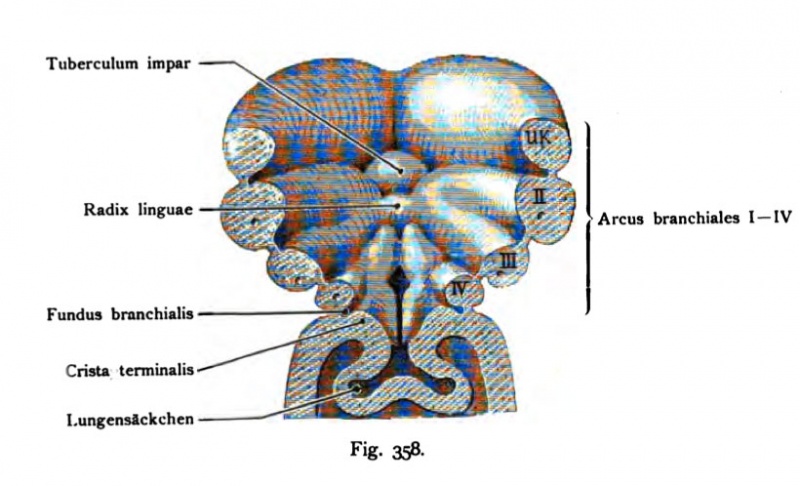
Fig. 358. Ventral wall of the foregut seen from the inside, also called the oral floor, that area from which the tongue, the larynx (voice box), thyroid (thyroid) and thymus arise
(After His.)
The dorsal portion of the head intestine along with the maxillary process and the brain tube removed. There are only so lateral and the front ends of the
Branchial arch visible and that the lower jaw extension of the First gill arch, the second or hyoid arch, then the III. and fourth branchial arch (Branchialbogen
called). In between lie the inner gill-pouches. The tuberculum impair, later front tongue is in the incision of the two Unlerkieferfortsätze. caudal the tuberculum impar to a connection string specifies the II and III. branchial arch, the basis of the lingual radix or root of the tongue is. This is followed by 357 in comparison to the figure had changed greatly changed furcula, which includes the installation of the epiglottis and the aryepiglottic folds. In the gap-like space between the medial ends of the furcula and Branchial arch develop the lateral thyroid gland.
- This text is a Google translate computer generated translation and may contain many errors.
Images from - Atlas of the Development of Man (Volume 2)
(Handatlas der entwicklungsgeschichte des menschen)
- Kollmann Atlas 2: Gastrointestinal | Respiratory | Urogenital | Cardiovascular | Neural | Integumentary | Smell | Vision | Hearing | Kollmann Atlas 1 | Kollmann Atlas 2 | Julius Kollmann
- Links: Julius Kollman | Atlas Vol.1 | Atlas Vol.2 | Embryology History
| Historic Disclaimer - information about historic embryology pages |
|---|
| Pages where the terms "Historic" (textbooks, papers, people, recommendations) appear on this site, and sections within pages where this disclaimer appears, indicate that the content and scientific understanding are specific to the time of publication. This means that while some scientific descriptions are still accurate, the terminology and interpretation of the developmental mechanisms reflect the understanding at the time of original publication and those of the preceding periods, these terms, interpretations and recommendations may not reflect our current scientific understanding. (More? Embryology History | Historic Embryology Papers) |
Reference
Kollmann JKE. Atlas of the Development of Man (Handatlas der entwicklungsgeschichte des menschen). (1907) Vol.1 and Vol. 2. Jena, Gustav Fischer. (1898).
Cite this page: Hill, M.A. (2024, April 27) Embryology Kollmann358.jpg. Retrieved from https://embryology.med.unsw.edu.au/embryology/index.php/File:Kollmann358.jpg
- © Dr Mark Hill 2024, UNSW Embryology ISBN: 978 0 7334 2609 4 - UNSW CRICOS Provider Code No. 00098G
Fig. 358. Ventrale Wand des Kopfdarms von innen gesehen, auch Mundboden genannt, jenes Gebiet, aus welchem die Zunge, der Kehlkopf (Larynx), Schilddrüse (Thyreoidea) und Thymus entstehen.
(Nach His.)
Der dorsale Abschnitt des Kopfdarms ist samt dem Oberkieferfortsatz und dem Hirnrohr entfernt. Es sind also nur die seithchen und vorderen Enden der Kiemenbogen sichtbar und zwar der Unterkieferfortsatz des I. Kiemenbogens, der II. oder Hyoidbogen, dann der III. und IV. Kiemenbogen (Branchialbogen genannt). Dazwischen liegen die inneren Kiementaschen. Das Tuberculum impar, später Vorderzunge, liegt im Einschnitt der beiden Unlerkieferfortsätze. Kaudal legt sich das Tuberculum impar an einen Verbindungsstrang des II. und III. Kiemenborcns, der die Grundlage der Radix linguae oder der Zungen wurzel darstellt. Darauf folgt die im Vergleich zu der Fig. 357 schon stark umgeänderte Furcula, welche die Anlage der Epiglottis und der Plicae aryepiglotticae umfaßt. In dem spaltartigen Raum zwischen Furcula und den medialen Enden der Kiemenbogen entwickeln sich die seitlichen Schilddrüsenanlagen.
File history
Click on a date/time to view the file as it appeared at that time.
| Date/Time | Thumbnail | Dimensions | User | Comment | |
|---|---|---|---|---|---|
| current | 13:00, 16 October 2011 |  | 820 × 498 (74 KB) | S8600021 (talk | contribs) | {{Kollmann1907}} |
You cannot overwrite this file.
File usage
The following page uses this file:
