File:Kollmann353.jpg
Original file (820 × 694 pixels, file size: 73 KB, MIME type: image/jpeg)
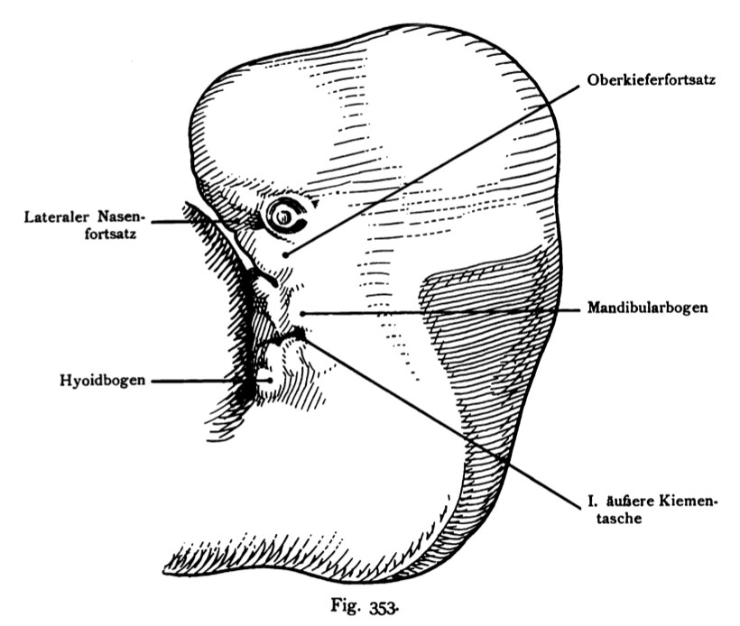
Fig. 353. Face of a Human Embryo of 12.6 mm, age 40 days
Left side of the profile.
(After Rabl.)
The mandibular arch is very sizeable. Behind him, the first outer Branchial pouch, ventral, lying close to the Herzwulst. Behind the pocket of the Hyoid arch. At the mandibular and hyoid arch at the cusps are noticeable, auricular hillocks (colliculi auricular) which used to build the ear.
- This text is a Google translate computer generated translation and may contain many errors.
Images from - Atlas of the Development of Man (Volume 2)
(Handatlas der entwicklungsgeschichte des menschen)
- Kollmann Atlas 2: Gastrointestinal | Respiratory | Urogenital | Cardiovascular | Neural | Integumentary | Smell | Vision | Hearing | Kollmann Atlas 1 | Kollmann Atlas 2 | Julius Kollmann
- Links: Julius Kollman | Atlas Vol.1 | Atlas Vol.2 | Embryology History
| Historic Disclaimer - information about historic embryology pages |
|---|
 |
Reference
Kollmann JKE. Atlas of the Development of Man (Handatlas der entwicklungsgeschichte des menschen). (1907) Vol.1 and Vol. 2. Jena, Gustav Fischer. (1898).
Cite this page: Hill, M.A. (2024, April 27) Embryology Kollmann353.jpg. Retrieved from https://embryology.med.unsw.edu.au/embryology/index.php/File:Kollmann353.jpg
- © Dr Mark Hill 2024, UNSW Embryology ISBN: 978 0 7334 2609 4 - UNSW CRICOS Provider Code No. 00098G
Fig. 353. Ausbildung des Gesichts von einem menschlichen Embryo
von 12,6 mm. Alter 40 Tage. Linke Seite des Profil.
(Nach Rabl.)
Der Mandibularbogen ist sehr ansehnlich. Hinter ihm die erste äußere Kiementasche, ventral, dicht am Herzwulst liegend. Hinter der Tasche der Hyoidbogen. Am Mandibular- und am Hyoidbogen sind Höcker bemerkbar, Aurikularhöcker, CoUiculi auriculares, die zum Aufbau der Ohrmuschel verwendet werden.
File history
Click on a date/time to view the file as it appeared at that time.
| Date/Time | Thumbnail | Dimensions | User | Comment | |
|---|---|---|---|---|---|
| current | 12:58, 16 October 2011 | | 820 × 694 (73 KB) | S8600021 (talk | contribs) | {{Kollmann1907}} |
You cannot overwrite this file.
File usage
The following page uses this file: