File:Kollmann352.jpg

Original file (873 × 687 pixels, file size: 66 KB, MIME type: image/jpeg)
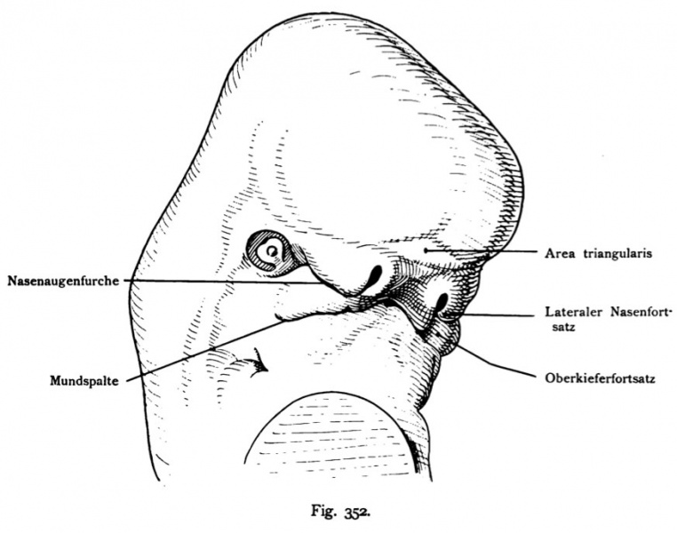
Fig. 352. Human embryo of 11.3 mm, 30-31 days
Head seen in half profile.
From the same embryo as shown in Figs 350 and 351. The head is up-oriented as in the adult. The triangular area on the forehead bulges more clearly in this view. Moreover, the furrow appears to mouth mouth and the palate through the shadows more noticeable. the Middle of the mandibular arch rises, making the mouth opening in the face handsomely arched against the forehead (see Fig 350 en face). the first Branchial pouch shows a considerable depth and beads as well as Fig. 351. The epithelium between the eyes and nose furrows shows the lacrimal drainage ways (Lacrimal canaliculi, lacrimal sac and duct) its course. She and the Wangenfiirche, the continuation of the mouth can in arrested development persist.
- This text is a Google translate computer generated translation and may contain many errors.
Images from - Atlas of the Development of Man (Volume 2)
(Handatlas der entwicklungsgeschichte des menschen)
- Kollmann Atlas 2: Gastrointestinal | Respiratory | Urogenital | Cardiovascular | Neural | Integumentary | Smell | Vision | Hearing | Kollmann Atlas 1 | Kollmann Atlas 2 | Julius Kollmann
- Links: Julius Kollman | Atlas Vol.1 | Atlas Vol.2 | Embryology History
| Historic Disclaimer - information about historic embryology pages |
|---|
 |
Reference
Kollmann JKE. Atlas of the Development of Man (Handatlas der entwicklungsgeschichte des menschen). (1907) Vol.1 and Vol. 2. Jena, Gustav Fischer. (1898).
Cite this page: Hill, M.A. (2024, April 27) Embryology Kollmann352.jpg. Retrieved from https://embryology.med.unsw.edu.au/embryology/index.php/File:Kollmann352.jpg
- © Dr Mark Hill 2024, UNSW Embryology ISBN: 978 0 7334 2609 4 - UNSW CRICOS Provider Code No. 00098G
Fig. 352. Menschlicher Embryo von 11,3 mm, 30—31 Tage. Kopf im Halbprofil gesehen.
Von demselben Embryo wie die Fig. 350 und 351. Der Kopf ist aufge- richtet wie bei dem Erwachsenen. Die Area triangularis an der Stirn wölbt sich bei dieser Ansicht deutlicher. Überdies erscheint die Furche zur Mund- öfFnung und zum Gaumen durch den Schlagschatten mehr bemerkbar. Die Mitte des Mandibularbogens erhebt sich, wodurch die Mundspalte im Gesicht ansehnlich gegen die Stirn gewölbt ist (vergl. Fig. 350 en face). Die erste Kiementasche zeigt eine ansehnliche Tiefe und ebenso Wülste wie Fig. 351. Die Augennasenftirche mit Epithel gefüllt, zeigt den ableitenden Tränenwegen (Canaliculi lacrimales, Saccus und Ductus lacrimalis) ihren Verlauf. Sie und die Wangenfiirche, die Fortsetzung der Mundspalte können bei Hemmungsbildungen persistieren.
File history
Click on a date/time to view the file as it appeared at that time.
| Date/Time | Thumbnail | Dimensions | User | Comment | |
|---|---|---|---|---|---|
| current | 12:58, 16 October 2011 | | 873 × 687 (66 KB) | S8600021 (talk | contribs) | {{Kollmann1907}} |
You cannot overwrite this file.
File usage
The following page uses this file: