File:Kollmann343.jpg

Original file (819 × 729 pixels, file size: 81 KB, MIME type: image/jpeg)
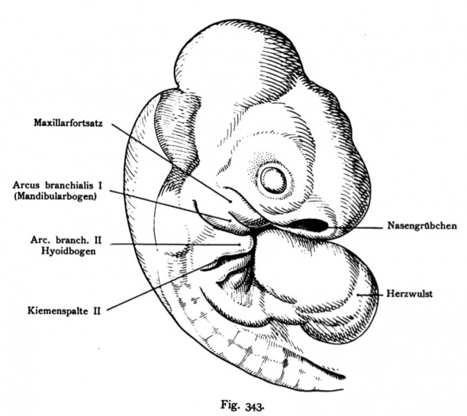
Fig. 343. Head of a Lizard Embryo (Sphenodon punctatum Hatteria)
Shortly before hatching, Norma lateralis to the many similarities to show the human embryo. Original image was a 20x magnification (see Fig 341)
(According to Schauinsland.)
Forward the heart lying close to head. The Nasengrübchen and the eye then follow the gill arch. The first is the gröfite of two departments, the maxillary and consisting of mandibular anlage, followed by the hyoid arch, which further branchial arch anschliefien. In all, five gill arches, separated by gill-pouches. The last two arc start (4 and 5) itself to lower into the depths. Are behind the arc formation proto-vertebrae (somites) noticeable.
- This text is a Google translate computer generated translation and may contain many errors.
Images from - Atlas of the Development of Man (Volume 2)
(Handatlas der entwicklungsgeschichte des menschen)
- Kollmann Atlas 2: Gastrointestinal | Respiratory | Urogenital | Cardiovascular | Neural | Integumentary | Smell | Vision | Hearing | Kollmann Atlas 1 | Kollmann Atlas 2 | Julius Kollmann
- Links: Julius Kollman | Atlas Vol.1 | Atlas Vol.2 | Embryology History
| Historic Disclaimer - information about historic embryology pages |
|---|
| Pages where the terms "Historic" (textbooks, papers, people, recommendations) appear on this site, and sections within pages where this disclaimer appears, indicate that the content and scientific understanding are specific to the time of publication. This means that while some scientific descriptions are still accurate, the terminology and interpretation of the developmental mechanisms reflect the understanding at the time of original publication and those of the preceding periods, these terms, interpretations and recommendations may not reflect our current scientific understanding. (More? Embryology History | Historic Embryology Papers) |
Reference
Kollmann JKE. Atlas of the Development of Man (Handatlas der entwicklungsgeschichte des menschen). (1907) Vol.1 and Vol. 2. Jena, Gustav Fischer. (1898).
Cite this page: Hill, M.A. (2024, April 27) Embryology Kollmann343.jpg. Retrieved from https://embryology.med.unsw.edu.au/embryology/index.php/File:Kollmann343.jpg
- © Dr Mark Hill 2024, UNSW Embryology ISBN: 978 0 7334 2609 4 - UNSW CRICOS Provider Code No. 00098G
Fig. 343. Kopf eines Eidechsenembryo (Sphenodon punctatum = Hatteria)
kurz vor dem Ausschlüpfen, Norma lateralis, um die mannigfachen Übereinstimmungen mit dem Menschenembryo zu zeigen. 20 mal vergr. (Vergl. Fig. 341)
(Nach Schauinsland.)
Vorn das Herz dicht am Kopf liegend. Dem Nasengrübchen und dem Auge folgen dann die Kiemenbogen. Der erste ist der gröfite, aus zwei Abteilungen, der Maxillar- und der Mandibularanlage bestehend, dann folgt der Hyoidbogen, dem sich weitere Kiemenbogen anschliefien. Im ganzen fünf Kiemenbogen, getrennt durch Kiementaschen. Die beiden letzten Bogen (4 und 5) beginnen sich schon in die Tiefe zu senken. Hinter den Bogenbildungen sind Proto-vertebrae (Urwirbel) bemerkbar.
File history
Click on a date/time to view the file as it appeared at that time.
| Date/Time | Thumbnail | Dimensions | User | Comment | |
|---|---|---|---|---|---|
| current | 12:55, 16 October 2011 |  | 819 × 729 (81 KB) | S8600021 (talk | contribs) | {{Kollmann1907}} |
You cannot overwrite this file.
File usage
The following 2 pages use this file:
