File:Kellicott 184.jpg

Original file (661 × 1,000 pixels, file size: 196 KB, MIME type: image/jpeg)
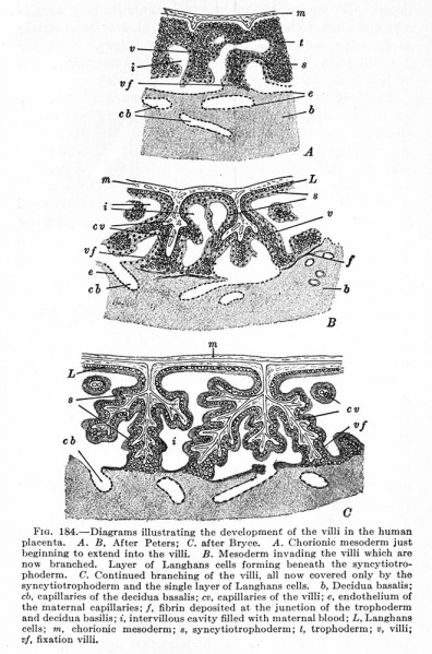
Fig. 184. Diagrams illustrating the development of the villi in the human placenta
A. B, After Peters; C. after Bryce.
A. Chorionic mesoderm just beginning to extend into the villi.
B. Mesoderm invading the villi which are now branched. Layer of Langhans cells forming beneath the syncytiotrophoderm.
C. Continued branching of the villi, all now covered only by the syncytiotrophoderm and the single layer of Langhans cells,
b, Decidua basalis; cb, capillaries of the decidua basalis; cv, capillaries of the villi; e, endothelium of the maternal capillaries; /, fibrin deposited at the junction of the trophoderm and decidua basilis; i, intervillous cavity filled with maternal blood; L, Langhans cells; m, chorionic mesoderm; s, syncytiotrophoderm; t, trophoderm; v, villi; vf, fixation villi.
| Historic Disclaimer - information about historic embryology pages |
|---|
| Pages where the terms "Historic" (textbooks, papers, people, recommendations) appear on this site, and sections within pages where this disclaimer appears, indicate that the content and scientific understanding are specific to the time of publication. This means that while some scientific descriptions are still accurate, the terminology and interpretation of the developmental mechanisms reflect the understanding at the time of original publication and those of the preceding periods, these terms, interpretations and recommendations may not reflect our current scientific understanding. (More? Embryology History | Historic Embryology Papers) |
Kellicott WE. Outlines of Chordate Development (1913) Henry Holt and Co., New York.
Cite this page: Hill, M.A. (2024, April 27) Embryology Kellicott 184.jpg. Retrieved from https://embryology.med.unsw.edu.au/embryology/index.php/File:Kellicott_184.jpg
- © Dr Mark Hill 2024, UNSW Embryology ISBN: 978 0 7334 2609 4 - UNSW CRICOS Provider Code No. 00098G
File history
Click on a date/time to view the file as it appeared at that time.
| Date/Time | Thumbnail | Dimensions | User | Comment | |
|---|---|---|---|---|---|
| current | 07:15, 28 August 2013 |  | 661 × 1,000 (196 KB) | Z8600021 (talk | contribs) | ==Fig. 184. Diagrams illustrating the development of the villi in the human placenta== A. B, After Peters; C. after Bryce. A. Chorionic mesoderm just beginning to extend into the villi. B. Mesoderm invading the villi which are now branched. Layer of L... |
You cannot overwrite this file.
File usage
The following 2 pages use this file:
