File:Kellicott 180.jpg
Original file (1,007 × 800 pixels, file size: 77 KB, MIME type: image/jpeg)
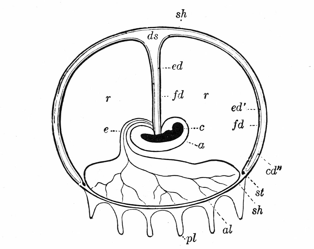
Fig. 180. Diagrammatic section through the fully formed blastodermic vesicle of the rabbit
Diagrammatic section through the fully formed blastodermic vesicle of the rabbit, showing the reduced yolk-sac. From Hertwig (Lehrbuch,.eJc.) after Bischoff.
o, Amnion; al, allantois; ds, yolk-sac; e, embryo; ed', ed f , ed" yolk-sac endoderm; fd, vascular layer (mesoderm) of yolk-sac; pi, villi; r, exoccelom; st, sinus terminalis; u, allantoic stalk.
| Historic Disclaimer - information about historic embryology pages |
|---|
 |
Kellicott WE. Outlines of Chordate Development (1913) Henry Holt and Co., New York.
Cite this page: Hill, M.A. (2024, April 27) Embryology Kellicott 180.jpg. Retrieved from https://embryology.med.unsw.edu.au/embryology/index.php/File:Kellicott_180.jpg
- © Dr Mark Hill 2024, UNSW Embryology ISBN: 978 0 7334 2609 4 - UNSW CRICOS Provider Code No. 00098G
File history
Click on a date/time to view the file as it appeared at that time.
| Date/Time | Thumbnail | Dimensions | User | Comment | |
|---|---|---|---|---|---|
| current | 16:44, 23 December 2013 | | 1,007 × 800 (77 KB) | Z8600021 (talk | contribs) | ==Fig. 180. == {{Outlines of Chordate Development footer}} Category:Rabbit |
You cannot overwrite this file.
File usage
The following 2 pages use this file: